The Understanding the Atom Series
Nuclear energy is playing a vital role in the life of every man, woman, and child in the United States today. In the years ahead it will affect increasingly all the peoples of the earth. It is essential that all Americans gain an understanding of this vital force if they are to discharge thoughtfully their responsibilities as citizens and if they are to realize fully the myriad benefits that nuclear energy offers them.
The United States Atomic Energy Commission provides this booklet to help you achieve such understanding.

Edward J. Brunenkant, Director Division of Technical Information
UNITED STATES ATOMIC ENERGY COMMISSION
by Walter E. Kisieleski and
Renato Baserga
United States Atomic Energy Commission
Division of Technical Information
Library of Congress Catalog Card Number: 66-61908
1966; 1967(Rev.)
The cover design portrays the inter-relationships suggested by the title of this booklet: On a trefoil symbolizing radiation are superimposed a dividing cell, a plant, an animal, and a double helix of a molecule of deoxyribonucleic acid, a material unique in and fundamental to all living things.
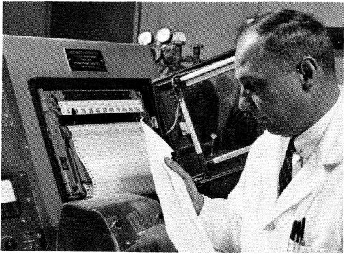
WALTER E. KISIELESKI is an Associate Scientist in the Division of Biology and Medicine of the Argonne National Laboratory. He was formerly associate professor of chemistry at Loyola University in Chicago. His undergraduate studies were at James Millikin University in Decatur, Illinois, and his graduate studies were at the University of Chicago. He received an Honorary Doctor of Science degree from James Millikin University in 1962. In 1958 he was a delegate to the Second Atoms for Peace Conference in Geneva, Switzerland. He was visiting lecturer in the department of biochemistry at the University of Oslo in Norway in 1963. Dr. Kisieleski is shown operating an automatic windowless strip counter that scans paper chromatograms and thus locates labeled substances.
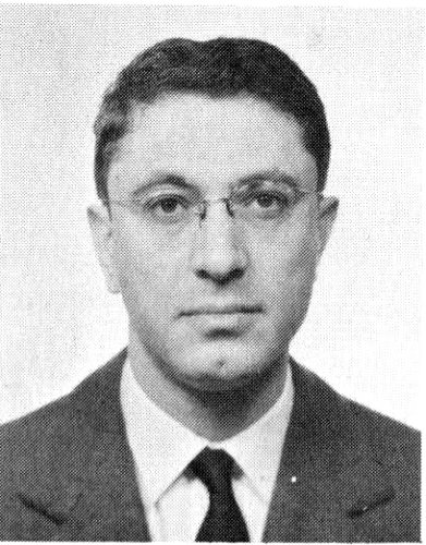
RENATO BASERGA was born in Milan, Italy, and received a medical degree from the University of Milan in 1949. He is presently research professor of pathology at the Fels Research Institute at Temple University Medical School in Philadelphia, and associate editor of the journal, Cancer Research. Formerly he was associate professor of pathology at Northwestern Medical School in Chicago, where he was the recipient of a Research Career Development Award from the National Institutes of Health.
By WALTER E. KISIELESKI
and RENATO BASERGA
Here and elsewhere we shall not obtain the best insight into things until we actually see them growing from the beginning.
Aristotle
The nature of life has excited the interest of human beings from the earliest times. Although it is still not known what life is, the characteristics that set living things apart from lifeless matter are well known. One feature common to all living things, from one-celled creatures to complex animals like man, is that they are all composed of microscopic units known as cells.
The cell is the smallest portion of any organism that exhibits the properties we associate with living material. In spite of the immense variety of sizes, shapes, and structures of living things, they all have this in common: They are composed of cells, and living cells contain similar components that operate in similar ways. One might say that life is a single process and that all living things operate on a single plan.
The past few years have been a time of rapid progress in our understanding of the mechanisms that control the function of living systems. This progress has been made possible by the development of new experimental techniques and by the perfection of instruments that detect what happens in the tiny world of molecules. Prominent among the methods that have contributed to the explosive growth 2 in our understanding of biology is the use of radioactive isotopes as laboratory tools.
In this booklet we shall attempt to give an account, in chemical terms, of the materials from which living matter is made and of some of the chemical reactions that underlie the manifestations and the maintenance of life. To accomplish this, we have chosen to describe three types of molecules that have become the basis of modern biology: deoxyribonucleic acid (DNA), ribonucleic acid (RNA), and proteins. We will show how radioactive isotopes can be used to pry into the innermost secrets of these substances. Before we can understand the function of these precious molecules, however, it will be necessary to review the structure of a cell and the physical nature of radioactive isotopes.
We have seen that all organisms are composed of essentially like parts, namely cells; that these cells are formed and grow in accordance with essentially the same laws; hence that these processes must everywhere result from the operation of the same forces.
Theodor Schwann
The cell theory, based on the concept that higher organisms consist of smaller units called cells, was formulated in 1838 by two German biologists, Mathias-Jacob Schleiden, a botanist, and Theodor Schwann, an anatomist. The theory had far-reaching effect upon the study of biological phenomena. It suggested that living things had a common basis of organization. Appreciation of its full significance, however, had to await more precise knowledge of the structure and activities of cells.
Some organisms,[1] for instance, amoebae, consist of a single cell each and are therefore called unicellular organisms. Higher animals are multicellular, containing aggregations of cells grouped into tissues and organs. A 3 man, for instance, consists of millions of many different cells performing a variety of different functions. Cells of higher animals differ vastly from one another in size, shape, and function; they are specialized cells.
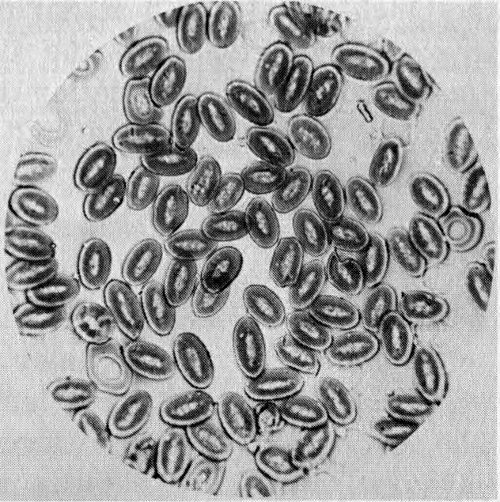
Figure 1 One of the earliest photographs of cells taken with a microscope. This photomicrograph shows cells in the blood of a pigeon. It was made by J. J. Woodward, U. S. Army surgeon, in 1871. Woodward had made the first cell micrograph (a graphic reproduction of the image of an object formed by a microscope) in 1866.
There is a remarkable similarity, moreover, in the molecular composition and metabolism[2] of all living things. This similarity has been taken to mean that life could have originated only once in the past and had a specific chemical composition on which its metabolic processes depended. This structure and metabolism were handed down to subsequent living things by reproduction, and all variations thereafter resulted from occasional mutation, or changes in the nature of the heredity-transmitting units. One of the most extraordinary of all the attributes of life is its ordered complexity, both in function and structure.
It is agreed among biologists that the functional manifestations of life include movement, respiration, growth, irritability (reaction to environmental changes), and reproduction and that these phenomena are therefore possessed by all cells. The first four of these can be grouped under a single word: metabolism. We can therefore say that living things have two common properties: metabolism and reproduction. Therefore, when we say we are studying life processes, we actually are studying the metabolism and reproduction of cells. Since metabolism is the sum of 4 the biochemical reactions taking place in a living organism, it properly belongs to the field of investigation of biochemists. Cell reproduction is the concern of both biochemists and morphologists[3] since it can be studied by either biochemical or morphological techniques.
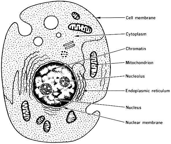
Figure 2 Generalized diagram of a cell, showing the organelles, or “little organs”, of its internal structure. The organelles that are labeled are important for this booklet.
The basic structure of a cell is shown in Figure 2. Each cell consists of a dense inner structure called the nucleus, which is surrounded by a less dense mass of cytoplasm. The nucleus is separated from the cytoplasm by a double envelope, called the nuclear membrane, which is peppered with perforations. The cytoplasm contains a network of membranes, which form the boundaries of countless canals 5 and vesicles (or pouches), and is laden with small bodies called ribosomes. This membranous network is called the endoplasmic reticulum and is distinct from the mitochondria, which are membranous organelles (little organs) structurally independent of other components of the cytoplasm. The outer coat of the cell is called the cell membrane, or plasma membrane, and forms the cell boundary.
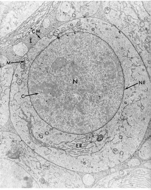
Figure 3 Electron micrograph of a primary spermatocyte cell of a grasshopper, showing the nucleus (N), endoplasmic reticulum (ER), mitochondrion (M), chromatin (C), nuclear membrane or nuclear envelope (NE), cell membrane (CM), and intercellular space (I). The magnification is about 25,000 times the actual size.
The nucleus, which in many cells is the largest and most central body, is of special importance. It contains a number of threadlike bodies, or chromosomes, that are the carriers of the cell’s heredity-controlling system. These contain granules of a material called chromatin, which is rich in a nucleic acid, DNA (deoxyribonucleic acid). The chromosomes usually are not readily seen in the nucleus except when the cell, along with its nucleus, is dividing. When the nucleus is not dividing, a spherical body, the nucleolus, can be seen. (In some nuclei there may be more than one.) When the nucleus is dividing, the nucleolus disappears.
Not all cells possess all these structures. For instance, the red cells of the blood do not have a nucleus, and in other cells the endoplasmic reticulum is at a minimum. The diagram (Figure 2) is valid for a great majority of the cells of higher organisms.
The cell structures shown in Figure 3 are visible with an electron microscope. They contain the chemical components of the cell. The chief classes of these constituents are the carbohydrates (sugars), the lipids (fats), the proteins, and the nucleic acids. However, a cell also contains water (about 70% of the cell weight is due to water) and several other organic and inorganic compounds, such as vitamins and minerals.
Carbohydrates serve mostly as foodstuff within the cell. They can be stored in several related forms. Further, they may serve a number of functions outside the cell, especially as structural units. In this way structure and function are correlated.
Lipids in the cell occur in a great variety of types: alcohols, fats, steroids, phospholipids, and aldehydes. They are found in all fractions of the cell. Their most important functions seem to be to form membranes and to give these membranes specific permeability. They are also important as stores of chemical energy, mostly in the form of neutral fats.
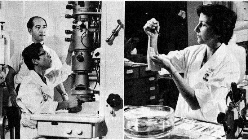
Figure 4 Scientists using an electron microscope (left) and an optical microscope (right) in fundamental biochemical research. Both instruments are important tools in studies of life processes.
The proteins occur in many cell structures and are of many kinds: Enzymes, the catalysts for the cell’s metabolic processes, are proteins, for instance. The nucleic acids are DNA and RNA (ribonucleic acid), which function together to manufacture the cell’s proteins. Since a large share of the remaining pages will be devoted to a discussion of proteins and nucleic acids, at this point we need only emphasize that these two types of materials are interrelated in their function and that both are essential.
It is not very fruitful to discuss whether proteins or nucleic acids are more important. That question is something like the one about the chicken and the egg. We cannot think of one without thinking of the other. Although our insight into the mutual dependence of these two materials has greatly increased in recent years and although we know the relation between them is a fundamental factor in such events as reproduction, mutation, and differentiation (or specialization) of cells, our understanding of their interplay is far from complete. Real understanding of the relation between them would give us insight into the essence of growth—both normal and abnormal—or, indeed, one could almost say, into the complexity of life itself.
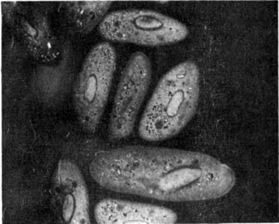
Figure 5 Photomicrograph of Paramecia, one-celled animals, magnified 1100 times. Many of the same structures that appear in Figure 3 can be seen here. This photo was taken with an “interference” microscope designed to permit continuous variation of contrast in the subject under study.
Practically all the DNA of most cells is concentrated in the nucleus. RNA, on the other hand, is distributed throughout the cell. Some RNA is present in the nucleus, but most of it is associated with minute particles in the cytoplasm known as microsomes, some of which are especially rich in RNA and are accordingly named ribosomes. These are much smaller particles than the mitochondria.
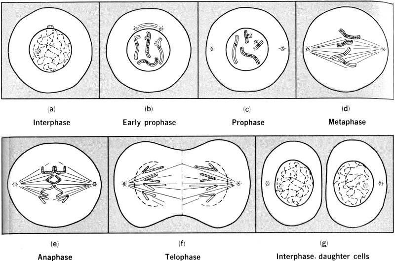
Figure 6 Stages of the mitotic cycle in a hypothetical cell with four chromosomes.
One of the most remarkable characteristics of cells is their ability to grow and divide. New cells come from preexisting cells. When a cell reaches a certain stage in its life, it divides into two parts. These parts, after another period of growth, can in turn divide. In this way plants and animals grow to their normal size and injured tissues are repaired. Cell division occurs when some of the contents of the cell have been doubled by replication, or copying (to be discussed later). The division of a cell results in two roughly equal new parts, the daughter cells. The process of cell division is known as mitosis and is diagrammed in Figure 6.
Mitosis is a continuous process; the following stages of the process are designated only for convenience. During interphase the cell is busy metabolizing, synthesizing new cellular materials, and preparing for self-duplication by synthesizing new chromosomes. In prophase the chromosomes, each now composed of two identical strands called chromatids, shorten by coiling, and the nucleolus and nuclear membrane disappear. During metaphase the chromosomes line up in one plane near the cell equator. At anaphase the sister chromatids of each chromosome separate, and each part moves toward the ends, or poles, of the cell. During telophase the chromosomes uncoil and return to invisibility; a new nucleus, nucleolus, and nuclear membrane are reconstituted at each end, and division of the cell body occurs between the new nuclei, forming the two new cells. Each daughter cell thereby receives a full 9 set of chromosomes, and, since the genes are in the chromosomes, each daughter cell has the same genetic complement.
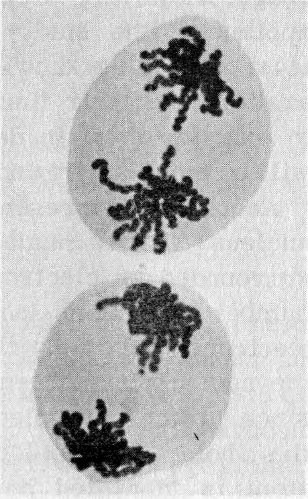
Figure 7 Photomicrograph of cells of the Trillium plant, which has five chromosomes, in anaphase. Note the duplicate sets of chromosomes moving to opposite poles of the cell.
All life processes use up energy and therefore require fuel. The mitochondria have a central role in the reactions by which the energy of sugars is supplied for cellular activity. The importance of this vital activity is obvious. In this booklet, however, we are concerned with the processes, involving nucleic acids and proteins, that can be described as making up “the gene-action system”. The gene-action system is the series of biochemical events that regulate and direct all life processes by “transcription” of the genetic “information” contained in molecules of DNA.
Man ... has found ways to amplify his senses ... and, with a variety of instruments and techniques, has added kinds of perception that were missing from his original endowment.
Glenn T. Seaborg
Practically everyone nowadays is to some extent familiar with the atomic structure of matter. Atomic energy, nuclear reactors, and radioisotopes are terms in everyday usage. However, to appreciate how radioisotopes can be applied to the study of life processes, we must have at least a working knowledge of their properties, their preparation, and their limitations. It is therefore appropriate to examine them in detail so that the succeeding chapters will be more easily understood.
According to present-day theory, an atom consists of a nucleus[4] that is made up of protons and neutrons[5] and is surrounded by electrons. In each atom there is an equal number of protons (positively charged) in the nucleus and electrons (negatively charged) moving concentrically around the nucleus; since neutrons have no electrical charge and since protons and electrons cancel each other’s charges, the whole atom is electrically neutral, or uncharged. Each atom is identified by an atomic number and an atomic weight. The atomic number of an element (for example, carbon, nitrogen, oxygen) is determined by the number of protons, or positive charges, carried by the nucleus (or by the number of electrons surrounding the nucleus, which is the same). The atomic weight is the weight of an atom as compared with that of the atom of carbon, which is taken as a standard. The weight, or mass, of an atom is due 11 chiefly to its protons and neutrons because the mass of its electrons is negligible.
Atoms of the same element, that is, atoms with the same number of protons and electrons, may vary slightly in mass because of having different numbers of neutrons. Since the chemical behavior of an element depends upon its electrons’ electrical charges, extra neutrons (which do not have an electrical charge) may affect the mass of an atom without disturbing its chemical properties. Atoms having the same atomic number but different atomic weights are called isotopes. For example, as shown in Figure 8, the isotope ¹H, or ordinary hydrogen, consists of a nucleus containing a proton (charge: +1; mass: 1) around which revolves an electron (charge: -1; mass: negligible); ²H, known as deuterium, contains an additional nuclear particle, a neutron (charge: 0; mass: 1); ³H, or tritium, contains two neutrons. Since the chemical behavior of an element depends upon the number of its electrons, these three atoms, although differing in weight, behave identically in chemical reactions. For convenience, the atomic weight is written as a superscript to the left of the element’s symbol. For instance ¹⁴C is the isotope of carbon with an atomic weight of 14 (ordinary carbon is the isotope with an atomic weight of 12, and it is written ¹²C).
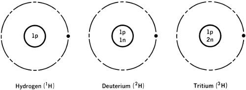
Figure 8 Isotopes of hydrogen.
Practically all elements have more than one isotope. There are two general classes of isotopes, stable and 12 radioactive. Stable isotopes have no distinguishing characteristic other than their mass; radioactive isotopes not only differ from their brothers in mass but also are characterized by unstable nuclei. When the nucleus of an atom is unstable, because of an unbalanced number of protons and neutrons, a redistribution occurs sooner or later, and the atom decomposes spontaneously and emits one of several kinds of radiations. Because of their common mode of action and effects on living organisms, these different kinds of radiations are known collectively as ionizing radiations.
All radioactive elements emit one or more of three types of penetrating (ionizing) rays. Alpha rays or particles are double-charged helium nuclei, ⁴He (atomic number: 2; mass: 4). They are emitted by many heavy radioactive elements, such as radium, uranium, and plutonium. Beta rays or particles can be either positive or negative. Negative beta particles are high-speed electrons and are emitted by many radioactive elements. Positive beta particles are positively charged electrons (positrons), have only a transitory existence, and are less common. Gamma rays are electromagnetic radiations, a term that also describes radiowaves, infrared rays, visible light, ultraviolet light, and X rays. Gamma rays are usually emitted after the emission of alpha or beta particles. In our studies of life processes, we are interested only in the radioactive isotopes that emit gamma rays or beta particles.
Radioactive isotopes occur as minor constituents in many natural materials, from which they can be concentrated by fractionation procedures. In a very limited number of cases, more significant amounts of a radioactive isotope, for example, radium or radioactive lead, can be found in nature. Most radioactive isotopes in use today, however, are prepared artificially by nuclear reactions. When a high-energy particle, such as a proton, a deuteron, an alpha particle, or a neutron, collides with an atom, a reaction takes place, leading to the formation of a new, unstable compound—a man-made radioactive isotope.
The great usefulness of radioactive isotopes, as we shall see later, is that they can be detected and identified by proper instruments. Biochemists have long recognized the desirability of “tagging” or “labeling” a molecule to permit tracing or keeping track of the “label” and consequently of the molecule as it moves through a reaction or process. Since the radiations emitted by radioactive isotopes can be detected and measured, we can readily follow a molecule tagged with a radioactive atom.
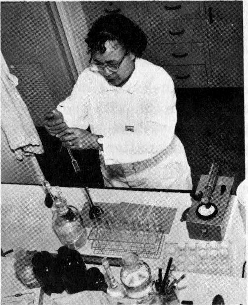
Figure 9 A laboratory technologist preparing dissolved biological materials as part of a study of the uptake of radioactive substances in living organisms. Note the radiation-detection instrument at right.
The earliest biochemical studies employing radioactive isotopes go back to 1924, when George de Hevesy used natural radioactive lead to investigate a biological process. It was only after World War II, however, when artificially made radioactive isotopes were readily available, that the technique of using isotopic tracers became popular.
In our investigations of life processes, we are especially interested in three radioactive isotopes: ³H, the hydrogen atom of mass 3; ¹⁴C, the atom of carbon with atomic weight 14; and ³²P, the atom of phosphorus with atomic weight 32. These radioactive isotopes are important because the corresponding stable isotopes of hydrogen, carbon, and phosphorus are present in practically all cellular components that are important in maintaining life. With the three radioactive isotopes, therefore, we can tag or label the molecules that participate in life processes.
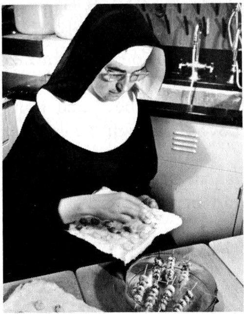
Figure 10 A visiting scientist at an AEC laboratory uses radioactive tritium (³H) to study the effect of radiation on bean chromosomes. The famous scientist, George de Hevesy, also used beans in conducting the first biological studies ever made with radioisotopes.
Hydrogen-3 is a weak beta emitter; that is, it emits beta particles with a very low energy (0.018 Mev[6]) and therefore with a very short range. Carbon-14 is also a weak beta emitter (0.154 Mev), although the beta particles emitted by ¹⁴C have a higher energy and therefore a longer range than those emitted by ³H. The beta particles emitted by phosphorus-32 are quite energetic (1.69 Mev) and have a longer range.
To biologists, then, the essential feature in the use of radioactive isotopes is the possibility of preparing “labeled” samples of any organic molecule involved in biological processes. With labeled samples it is possible to distinguish the behavior and keep track of the course of molecules involved in a particular biological function.
In this capacity the isotope may be likened to a dynamic and revolutionary type of “atomic microscope”, which can actually be incorporated into a living process or a specific cell. Just as a real microscope permits examination of the structural details of cells, isotopes permit examination of the chemical activities of molecules, atoms, and ions as they react within cells. (Neither optical nor electron microscopes are powerful enough for us to see anything as small as a molecule clearly.)
Here, surely, is the prime substance of life itself.
Isaac Asimov
The many characteristic features of each living species, its complex architecture, its particular behavior patterns, the ingenious modifications of structure and function that enable it to compete and survive—all these must pass, figuratively speaking, through the eye of an ultramicroscopic needle before they are brought together as a new, individual organism. The thread that passes through the eye of this needle is a strand of the filamentous molecule, deoxyribonucleic acid (DNA). Let us now outline the research that led to these conclusions.
One of the fundamental laws of modern biology—which states that the DNA content of somatic cells is constant for any given species—was first set forth in a research report of 1948. This finding means that in any given species, such as a mouse or a man, all cells except the germinal cells contain the same amount of DNA. Germinal cells, that is, the sperm cells of the male semen and the female egg, contain exactly half the amount of DNA of the somatic cells. This must be the case, since DNA is the hereditary material, and each individual’s heredity is shaped half by his father and half by his mother. One ten-trillionth of an ounce of DNA from a father and one ten-trillionth of an ounce of DNA from the mother together contain all the specifications to produce a new human being.
A large amount of DNA must be manufactured by an individual organism as it develops from a fertilized egg (one single cell) to an adult containing several million cells. For instance, a mouse cell contains about 7 picograms of DNA (one picogram is one millionth of a microgram, 16 or one millionth of one millionth of a gram). A whole mouse contains in its body approximately 25 milligrams (25 thousandths of a gram) of DNA, and all this DNA was synthesized by the cells as the mouse grew to adulthood. Since the amount of DNA per cell remains constant and since each cell divides into two cells, it is apparent that each new cell receives the amount of DNA characteristic of that species.
Once we realize that a cell that is making new DNA (as most cells do) must divide to keep the amount of DNA per cell constant, it follows that a cell that is making DNA is one that is soon destined to divide. If we can now mark newly made DNA with a radioactive isotope, we can actually mark and thus identify cells that are preparing to divide. The task can be divided into two parts: (1) to label the newly made DNA and (2) to detect the newly made, labeled DNA.
Figure 11 is a diagram showing the essential structure of the large DNA molecule. According to the Watson-Crick model,[7] the molecule consists of two strands of smaller molecules twisted around each other to form a double helix. Each strand consists of a sequence of the smaller molecules linked linearly to each other. These smaller molecules are called nucleotides, and each consists of three still smaller molecules, a sugar (deoxyribose), phosphoric acid, and a nitrogen base. Each nucleotide and its nearest neighbor are linked together (between the sugar of one and the phosphoric acid of the neighbor). This leaves the nitrogen base free to attach itself, through hydrogen bonding, to another nitrogen base in the opposite strand of the helix.
In the DNA of higher organisms, there are only four types of nitrogen bases: adenine, guanine, thymine, and cytosine. Adenine in either strand of the helix pairs only with thymine in the opposite strand, and vice versa, and guanine pairs only with cytosine, and vice versa, so that 17 each strand is complementary in structure to the other strand (see Figure 12). The full structure resembles a long twisted ladder, with the sugar and phosphate molecules of the nucleotides forming the uprights and the linked nitrogen bases forming the rungs. Each upright strand is essentially a mirror image of the other, although the two ends of any one rung are dissimilar.

Figure 11 Diagrammatic structure of the DNA molecule as proposed by the Watson-Crick model.
When DNA is replicated, or copied, as the organism grows, the two nucleotide strands separate from each other by disjoining the rungs at the point where the bases meet, and each strand then makes a new and similarly complementary strand. The result is two double-stranded DNA molecules, each of which is identical to the parent molecule and contains the same genetic material. When the cell divides, each of the two daughter cells gets one of the new double strands; each new cell thus always has the same amount of DNA and the same genetic material as the parent cell.
(All that has been said so far about DNA replication depends upon an assumption that the DNA molecule is in 18 some way untwisted to allow separation of two helical strands, but there is no compelling reason to believe that such an untwisting does indeed take place, nor do we know, if the untwisting does take place, how it is accomplished. Much that has been said in the last few paragraphs is therefore purely speculative. It is, however, based on sound observation and is a more logical explanation than others that have been advanced.)
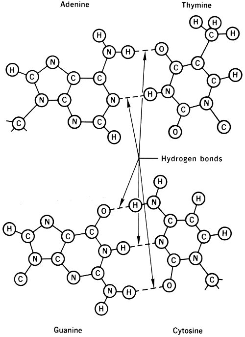
Figure 12 The pairing of the nucleotide bases that make up DNA.
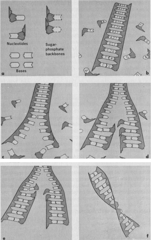
Figure 13 The DNA molecule and how it replicates. (a) The constituent submolecules. (b) Assembly of subunits in complete DNA molecule. (c) “Unzipping” of the double nucleotide strand. (d) and (e) The forming of a new strand by each individual strand. (f) DNA molecule in twisted double-strand configuration.
Adapted from Viruses and the Nature of Life, Wendell M. Stanley and Evans C. Valens, E. P. Dutton & Co., Inc., 1961, with permission.
Of the four bases in DNA, three are also found in the other nucleic acid, RNA; but the fourth, thymine, is found only in DNA. Therefore, if thymine could be labeled and introduced into a number of cells, including a cell in which DNA is being formed, we would specifically label the newly synthesized DNA, since neither the old DNA nor the RNA would make use of the thymine. We could in this way mark cells preparing to divide. (Actually, thymine itself is not taken up in mammalian cells, but its nucleoside is. A nucleoside is the base plus the sugar, or, in other words, the nucleotide minus the phosphoric acid.) The nucleoside of thymine is called thymidine, and we say that thymidine is a specific component of DNA and can be used, both in laboratory studies and in living organisms, for labeling DNA.
Thymidine labeled with radioactive compounds is available as ¹⁴C-thymidine (thymidine with a stable carbon atom replaced by a radioactive carbon atom) and as ³H-thymidine (thymidine in which a stable hydrogen atom has been replaced by tritium). Thus, when cells actively making DNA are exposed to radioactive thymidine, they incorporate it, and the DNA becomes radioactive.
We have thus found a way to complete the first part of the task, the labeling of new DNA. We still must find out how to distinguish labeled DNA among the many components of the cell. We might do it with a system based on measuring the amount of radioactivity incorporated into the DNA of cells exposed to radioactive thymidine, as an approximation of the frequency of cell division in the group of cells. However, a better method for studying cells synthesizing DNA, and thus preparing to divide, is the use of high-resolution autoradiography.
Autoradiography is based on the same principle as photography. Just as photons of light impinging on a photographic emulsion produce an image, so do beta particles (or alpha particles) emitted by decomposing radioactive atoms. A photographic emulsion is a suspension of 21 crystals of a silver halide (usually silver bromide) embedded in gelatin. When crystals of silver bromide are struck by beta particles, the silver atoms are ionized and form a latent image, so called because it is invisible to our eyes. After the emulsion is developed and fixed, each little aggregate of reduced silver atoms becomes a visible black speck on the emulsion. The distribution and combination of the specks make up the photographic image (see Figure 14). In ordinary photography such an image is a negative, which has to be converted into the positive photograph by printing. In autoradiography we are satisfied to look at the negative image since the clusters of developed silver atoms, appearing under a light microscope as black dots, supply all the information we need.
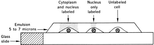
Figure 14 Schematic diagram of a radioautograph.
The distinction of having made the first autoradiograph belongs to the French physicist, Antoine Henri Becquerel; and to another Frenchman, A. Lacassagne, goes the credit for having introduced this technique into biological studies. Lacassagne used autoradiography to study distribution of radioactive polonium in animal organs. After World War II, when radioactive isotopes were first available in appreciable quantities, autoradiography was further perfected through the efforts of such scientists as C. P. Leblond in Canada, S. R. Pelc in England, and P. R. Fitzgerald in the United States.
Today autoradiography is sufficiently precise to locate radioactively labeled substances in individual cells and even in chromosomes and other structures within the cell. Two conditions must be met to achieve this high resolution: (1) The radiation from the radioactive element in the cells 22 must be of very short range. (2) The cells must remain in close contact with the photographic emulsion throughout the various experimental manipulations. When these conditions are met, the black dots will appear in the emulsion directly above the cell or cell part from which the radiation came (see Figure 14).
Shortness of range is satisfied by use of tritium, since its beta particles travel only about 1 micron (one thousandth of a millimeter) and the diameters of mammalian cells range from 15 to 40 or more microns. A mammalian-cell nucleus is at least 7 to 8 microns in diameter.
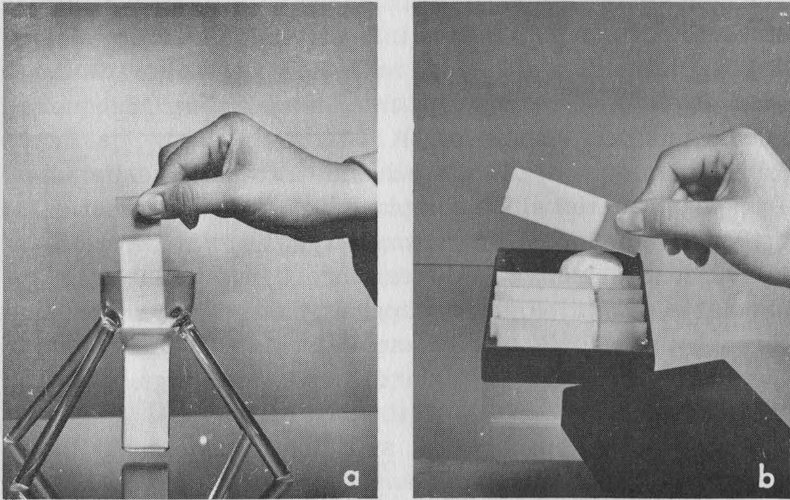
Figure 15 Cells being prepared for autoradiography. (a) Cells being coated with a photographic emulsion. (b) Coated cells being exposed to produce a latent image.
The condition of close contact between cells and emulsion is achieved by the technique of dip-coating autoradiography. In this process the glass slide on which the cells are carried is dipped into a melted photographic emulsion (see Figure 15a), a thin film of which clings to the slide. After it has been dried, the slide is placed in a lighttight box and kept in a refrigerator for the desired period of exposure, usually several days or weeks. During this period disintegrating radioactive atoms within the cells continue to emit beta particles, which, in turn, produce a latent image in the overlying emulsion. After the exposure is 23 complete, the slide is developed and fixed like a photographic plate, and a stain is applied which penetrates the emulsion so that the outlines of the cells and their internal structures can be seen. The fixing process removes all silver bromide that has not been ionized so that the emulsion is reduced to a thin, transparent film of gelatin covering the stained cells and containing only the clusters of silver grains that were struck by the beta particles.
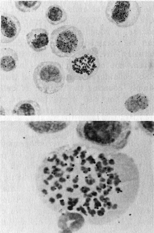
Figure 16 Radioautographs of tumor cells. Above, tumor cells and blood cells. Below, magnification of tumor cells.
When the finished autoradiograph is examined under the microscope, it will look like the radioautographs of tumor cells in Figure 16. In the upper micrograph the tumor cells are the larger ones and the smaller ones are blood cells. The dense structures in the center of the tumor cells are nuclei. The cells were exposed to tritium-labeled thymidine, and those synthesizing DNA at the time of exposure took up the thymidine and became radioactive. They can be identified by the black dots overlying the nuclei; the dots are the aggregates of silver grains struck by the beta particles.
Notice that only the nuclei contain radioactivity; the reason for this is that radioactive thymidine is incorporated only into DNA localized in the nuclei of cells. This picture identifies not only the cells that were making DNA at the time the label was administered but also the cells that were destined to divide in the immediate future, since cells synthesize DNA in preparation for cell division.
If we want to compare two populations of cells to find out which is proliferating (dividing) more actively, counting the fraction of cells labeled will give the number of cells synthesizing DNA in preparation for cell division. Of course, a rough approximation of the proliferating activity can be obtained by simply counting the number of cells actually dividing. But with tritiated thymidine we can obtain not only much more accurate measurements but also considerable information that cannot be obtained by simply counting the number of cells in mitosis. We shall discuss the cell cycle later on, but for the moment we should emphasize that much of our knowledge of the cell cycle stems from the use of high-resolution autoradiography.
It is clear that autoradiography enables us to find out which cells are dividing in a cell population and how many of them do so. For instance, in a given tissue or organ, not all cells are capable of dividing into two daughter cells. In the epidermis, which is the thin outer layer of the skin, only cells in the deepest portion can divide. The other cells, although originating from cells in the deep layer, have lost the capacity to divide, and eventually die without further division. If we take a bit of skin, expose it to tritiated thymidine, and determine the amount of radioactivity incorporated into the skin cells’ DNA, we obtain a fair measurement of the amount of DNA being synthesized. However, this purely biochemical investigation cannot possibly give any information on which specific cells are synthesizing DNA. For this, autoradiography provides the information we need.
Mathematicians are like Frenchmen: whatever you say to them they translate into their own language, and forthwith it is something entirely different.
Wolfgang von Goethe
We have mentioned previously that there are two main types of nucleic acids: DNA, the genetic material itself, and RNA, the molecule that translates the genetic message from DNA into terms the cell can use as “instructions” for making protein. Cells differ from each other on the basis of kinds of proteins they contain, and, since differences among cells determine differences among organisms, it follows that differences in the composition of DNA serve to explain the variety in living organisms populating the world. However, if differences between two organisms can be explained by differences in the chemical composition of their respective DNA’s, how can we explain differences between cells of the same organism? How can we explain that cells of the human pancreas secrete insulin, whereas other cells in man produce no insulin? Or how can we explain that certain cells make bone and others make fat? If indeed all cells in the same organism contain the same amount and kind of DNA (since all DNA in an organism derives from the duplication of the DNA of the fertilized egg cell and its descendants), it would seem, at first glance, that DNA is not the molecule responsible for differences among the cells. The clarification of this apparent contradiction is found in the remarkable properties of the other nucleic acid, the translator molecule, RNA.
In the first place, there are at least three different kinds of RNA. The largest quantity is a special kind called ribosomal RNA, or r-RNA. It is found in close conjunction with proteins and makes up the structural frame upon which the protein-synthesizing machinery is built. The r-RNA and the proteins to which it is firmly bound form 26 the ribosomes, the RNA-rich microsomes that are attached to the endoplasmic reticulum. Proteins are synthesized on ribosomes. We shall see later what determines the differences among proteins and how these differences are dictated directly by RNA and indirectly by DNA.
Besides r-RNA, there is a kind of RNA called soluble RNA, or transfer RNA, or s-RNA. It combines with r-RNA to complete the sequence of events that synthesizes the proteins. A bond between r-RNA and s-RNA is established by a third RNA molecule called messenger RNA, or template RNA, or m-RNA. This m-RNA molecule is truly the messenger that carries the genetic message from DNA to the protein-synthesizing apparatus.
Dr. Michael Shimkin, a Temple University scientist, in his analogy has compared the DNA → RNA → protein sequence to the activities of a newspaper staff. DNA is the editor; m-RNA molecules are copyboys who carry the editorials to the typesetters, the r-RNA and s-RNA, who then take the “letters” of nucleic acid and set them into slots in accordance with the editor’s directions. There are also workers who melt down outworn letters and still other workers who make new letters for further use; these are the enzymes, special kinds of proteins. If we wish to continue the analogy, we may say that each kind of cell in the organism has a different subeditor, who writes that cell’s own editorial. Actually we might say that all cells have the same board of editors in common, but only one editor functions in any given type of cell. In biological terms this means that only a portion of all the cellular DNA is active in each cell.
The active DNA is the DNA that makes m-RNA that will carry instructions to the protein-synthesizing machinery of that type of cell. Cells of the same organism therefore differ from each other on the basis of the segment of DNA that is active in making m-RNA. Let us now see how we can use radioactive isotopes to investigate the synthesis of RNA.
RNA synthesis is investigated with radioactive tracers in the same way as DNA synthesis. If we can mark, with a 27 radioactive atom, a small molecule that is incorporated into newly formed RNA, we can then trace the course of the labeled RNA molecule with a radiation-detection device. DNA had one advantage in this regard—the fact that one compound, thymidine, was a precursor of DNA, a specific material that could be incorporated only into DNA. We do not know similar specific precursors of RNA. But we know several precursors that are predominantly incorporated into RNA; the most common of these are the nucleosides adenine, cytidine, and uridine, and the smaller molecule, orotic acid. All these precursors can be labeled with either ³H or ¹⁴C, and their incorporation into RNA can be measured.
As in DNA synthesis, we can use autoradiography to follow the incorporation of precursors into RNA. By proper treatment of the tissues, we can make sure that all the radioactivity visible by autoradiography is due to labeled RNA, even though some of the precursor also enters DNA molecules. Even so, the kind of information obtained from autoradiographs of tissues exposed to RNA precursors is different from that obtained with DNA precursors. The advantage of high-resolution autoradiography in DNA studies is the possibility of identifying particular cells that are synthesizing nucleic acid. This advantage is apparently lost in the case of RNA. The reason is that, at any given time, only a few cells are making DNA, whereas practically all cells are synthesizing RNA constantly. The only exceptions are cells in the midpoint of mitosis. At the beginning (prophase) and at the end of cell division (telophase), RNA is synthesized. If we want a quantitative measurement of RNA synthesis, other methods, to be examined presently, are considerably more precise. But autoradiography can still give us valuable information.
If we look at cells soon after they have been exposed to an RNA precursor, we find that the radioactivity detectable by autoradiography is only in the nuclei of the cells. No 28 radioactivity can be detected in the cytoplasm, although we know that the cytoplasm of living cells contains large amounts of r-RNA and s-RNA. One or two hours later, however, radioactive RNA appears in the cytoplasm as well as in the nucleus. What autoradiography is telling us is that RNA is made in the nucleus and then is slowly transferred to the cytoplasm.
Autoradiography cannot tell us whether the RNA that has been newly synthesized in the nuclei of cells is m-RNA, s-RNA, or r-RNA. The methods necessary to make this distinction are based on the chemical fractionation of the tissue, isolation of RNA, determination of its amount by quantitative analysis, and determination of the amount of radioactivity by physical methods. Let us examine these steps separately.
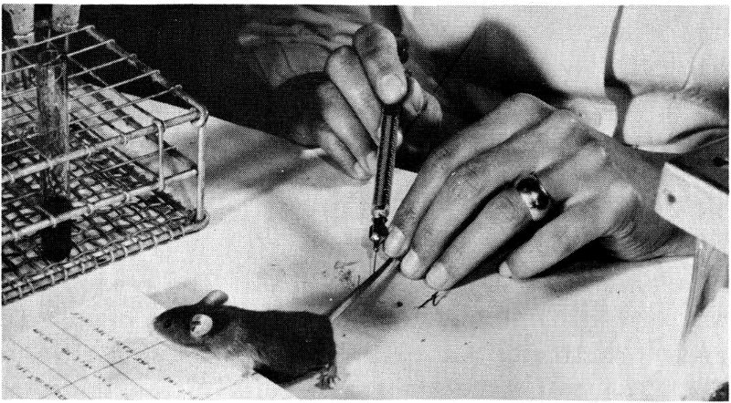
Figure 17 Injecting a mouse with a radioactive solution.
Chemical Fractionation of Tissue After an animal has been injected with a radioactive precursor of RNA, some of it will be incorporated into DNA as well as into RNA (remember that the precursors of RNA lack specificity), and part of the precursor will be broken down into smaller molecules. The injected animal can be sacrificed, and an organ or another tissue, for instance, the liver, can be removed. Then the liver is homogenized, that is, ground to a pulp with a modern version of the mortar and pestle. The homogenate (pulp) is treated with cold (weak) acid. Proteins and nucleic acids are insoluble in cold acids and 29 therefore precipitate to the bottom of the test tube. All molecules that are soluble in a cold acid are left in the supernatant (the remaining liquid); among these are small molecules, like those of the RNA precursor. The precipitate (the solid material that settles to the bottom), now containing proteins and nucleic acids, is then treated with a strong alkali, for instance, sodium hydroxide. Alkali will digest RNA into smaller molecules but does not affect DNA. If we now add acid to the solution, DNA, being insoluble in acid, will precipitate again; RNA, having been broken down into small molecules, will remain in the supernatant. DNA can then be extracted from the precipitate by boiling in strong acid. Proteins from the tissue remain in the final residue.
We have now fractionated the tissue into four portions: the acid-soluble fraction (containing small molecules), RNA, DNA, and proteins. (The cell’s lipids and sugars come out during alcohol rinses between the weak acid and the alkali steps.) Chemical analysis allows us to measure precisely the amount of RNA or DNA in its respective fraction and therefore in the tissue or organ. The amount of radioactivity in the RNA fraction can then be determined by a technique known as liquid scintillation counting.
Liquid Scintillation Counting Liquid scintillation counting is the preferred method for the measurement of low-energy beta-emitting radioisotopes commonly used in cell-fractionation studies (see Figure 18). It is convenient, sensitive, and rapid for routine measurement of radiation in hydrocarbons, other organic compounds, and aqueous solutions containing such isotopes as ³H, ¹⁴C, and ³²P.
Liquid scintillation solutions share with other scintillating materials the property of converting into visible light the energy deposited in them by ionizing radiation. In theory, if a sample of a beta emitter is dissolved in a liquid scintillator solution, every beta particle emitted will be absorbed completely because the range of penetration of beta particles in liquids is quite short (ranging from 0.008 millimicron for ³H to 7.9 millimicrons for ³²P in a medium of unit density). The kinetic energy of the beta particles is largely used up in the ionization and excitation of the most abundant molecular species present, the 30 solvent in which the scintillating material was dissolved. A fraction of the energy thus expended by each beta particle is transferred from excited solvent molecules to scintillator molecules; thus the electrons in the atoms of the scintillator molecules are raised to an excited (higher energy) state. When these electrons return to the ground, or unexcited, state, a fraction of them emit a photon of light. Thus each beta particle produces a burst of photons.
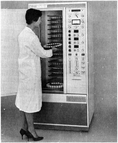
Figure 18 Technician placing a tray of samples in a liquid scintillation counter. The radioactivity of each sample is recorded as the trays revolve.
If a vessel containing the liquid scintillator and the radioactive sample is placed near a suitably sensitive instrument known as a photomultiplier tube, each burst of scintillator photons activates this device and causes it to release a burst of photoelectrons. Each burst of photoelectrons is multiplied successively in a series of electronic steps; as a result, there is a suitably large electrical-output pulse to be recorded.
One of the principal advantages of the liquid scintillation method is the ease of sample preparation. We need only transfer a known volume of a liquid sample or weigh a given mass of a solid sample into a sample bottle, add a known amount of the liquid scintillator solution, and stir until there is a homogeneous solution. Samples thus prepared are placed in a refrigerated counting apparatus. 31 After a short waiting period to allow time for the samples to cool and for a natural, short-lived phosphorescence (due to exposure to room light) to subside, the samples are ready to be measured.
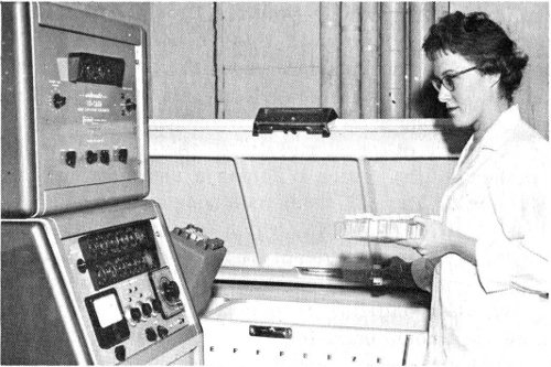
Figure 19 Placing radioactive samples in a refrigerated unit for liquid scintillation counting.
One disadvantage of liquid scintillation counting is that different compounds show different degrees of quenching (loss of emitted photons), and the effect must be checked for each class of compounds in each concentration range. This checking is usually done with an internal standard technique, the sample being counted before and after a standard, or known, emitter is added.
Another difficulty is that the best scintillating solvents are not the best chemical solvents for most biological materials. The solubility problem is also aggravated by the low temperatures at which liquid scintillation counters are usually operated for more effective instrument performance.
With the method we have described, we can obtain a fairly accurate idea of the rate of RNA synthesis in a given tissue. There are other things we would like to know about RNA. The first of these is the kind of RNA being synthesized. During alkaline digestion all kinds of RNA are broken down into their component nucleotides; we must therefore use other methods if we wish to know the kind of 32 RNA in which the radioactivity of the precursor has been incorporated.
Isolation of RNA Native RNA, that is, RNA not broken down into its smaller constituents, can be obtained in a variety of ways, but the most popular one makes use of phenol extraction, which removes DNA and proteins and leaves RNA in solution. If this phenol-purified RNA is dissolved in a concentrated sugar solution and spun in a centrifuge at a very high velocity, it will separate into three major components. These components separate because they have different molecular weights, and the larger the molecule, the faster it forms a sediment in the centrifugal field. Two of these components are s-RNA, the lightest of all, and r-RNA, which is divided into two subfractions. We can also identify a third component, m-RNA, with the centrifuge system but only with some difficulty and only after labeling it with a radioactive precursor, because the amount of m-RNA in a cell is very small.
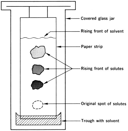
Figure 20 Diagram of ascending paper chromatography.
Quantitative Analysis Another important feature of RNA (or DNA, for that matter) is its base composition, that is, the percentage of each of the nucleotides that make it up. 33 The four bases that, with ribose and phosphoric acid, comprise the RNA molecule are guanine, adenine, cytosine, and uracil. It will be noted that three of the four—guanine, adenine, and cytosine—are the same as those in DNA, but thymidine in DNA has been replaced by another base, uracil. To determine the percentage of each base in a given RNA molecule, we must digest RNA with alkali to produce mononucleotides, which are smaller molecules, each consisting of a base, ribose, and phosphoric acid. We can now separate the four nucleotides by using paper chromatography (see Figure 20).
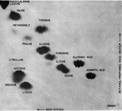
Figure 21 A paper chromatography showing separation of amino acids in two directions. Radioactivity in samples then produced this record by radioautography.
In this technique a mixture of compounds is deposited on the edge of a special type of paper. This edge is then immersed in a solvent that slowly permeates the paper (at a constant speed) by capillary action. As the solvent moves from the immersed edge toward the other edge, which is hanging freely, it carries the mixture of nucleotides with 34 it. Each of the compounds in the mixture travels at a different speed, however; thus, as the solvent front moves along the paper, the dissolved compounds are separated from each other and appear as distinct spots on the paper. To locate the nucleotides on the paper and to determine the percentage composition, we can use a chromatogram scanner, a device that scans the paper chromatograms, measures the radiation from them, and thus locates the labeled substances (see Figure 22).
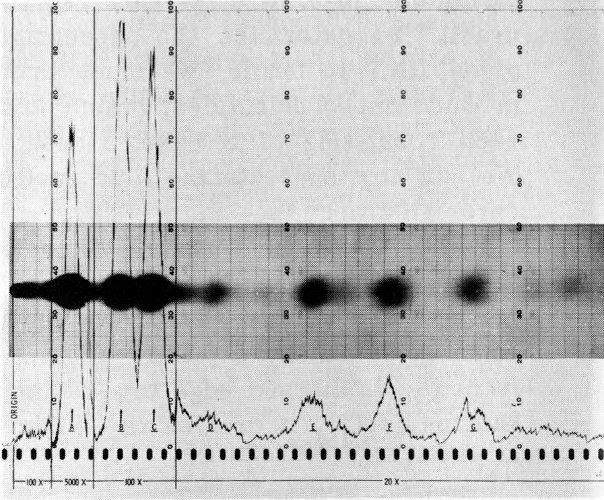
Figure 22 Recording of radioactivity in a sample by radioautography and paper chromatography. The peaks of the trace prepared by a chromatogram scanner coincide with the areas of separated components on the same chromatogram, as revealed by radioautography. The radioautograph is superimposed on the chromatogram recording.
Another technique used to separate the nucleotides of RNA is column chromatography. In this method mixtures of nucleotides are separated as they pass down a column of chemicals (see Figure 23).
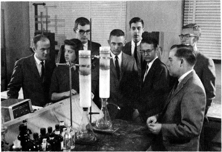
Figure 23 Students visiting Argonne National Laboratory listen to a scientist explain the column chromatography process, in which mixtures of nucleotides are separated as they pass down a column of chemicals.
We have now learned how to use radioisotopes to investigate the synthesis of RNA, the molecule that translates the DNA message into the language of proteins. Let us now see what we can learn about the synthesis and function of proteins.
If a man will begin with certainties he shall end in doubts; but if he will be content to begin with doubts he shall end in certainties.
Francis Bacon
Proteins occupy a central position in the structure and functioning of living matter and are intimately connected with all the metabolic reactions that maintain life. Some proteins serve as structural elements of the body, for instance, hair, wool, and the scleroproteins of bone and collagen, the latter an important constituent of connective tissue. Other proteins are enzymes, which are extremely important since they regulate all metabolic reactions. Most of the proteins in the tissues of actively functioning organs, such as the liver and the kidney, are enzymes. Other proteins participate in muscular contraction, and still others are hormones or oxygen carriers. Special proteins called histones are associated with gene function, and the antibodies that an organism produces to defend itself from bacteria are also proteins.
The differences in proteins, especially in enzymes, account for differences among cells. It is now appropriate to ask what makes one protein different from another. We know that the structure of a protein depends upon several factors, such as the molecular weight. But the main differences among proteins depend upon the sequence, or order, of the amino acids that are linked together in the protein molecules.
Amino acids are the fundamental structural units of proteins. There are 20 amino acids found frequently in mammalian proteins, and these molecules may be linked to one another to form a chain called a polypeptide chain. The structure of a protein then depends on: (1) the quantity of each amino acid present; (2) the sequence of amino acids in the polypeptide chain; (3) the length of the polypeptide chain, that is, the molecular weight; and (4) the folding and the side (nonlinear) arrangement of the polypeptide chain molecules, that is, the secondary and tertiary structures.
How can we investigate protein synthesis by using radioactive isotopes? Since proteins are made up of amino acids, the logical conclusion, after what we have learned about DNA synthesis and RNA synthesis, is that the best way would be to mark an amino acid and follow its incorporation into a molecule of protein. We could label a mixture of several amino acids, but, for the sake of clarity, we will describe the incorporation of a single labeled amino acid.
Suppose we have the amino acid leucine labeled with ¹⁴C and we inject a solution containing it into an experimental animal. Since leucine is incorporated into proteins, if we isolate the proteins and determine both the amount of proteins and the amount of radioactivity, we can measure fairly accurately the rate of protein synthesis. Autoradiography, by the way, is of little help in studying most protein synthesis because all cells are always synthesizing proteins and so are all labeled after a single exposure to a radioactive amino acid. With RNA precursors autoradiography at least told us where RNA was being made, but with amino acids we do not even get this information because proteins are synthesized both in the nucleus and in the cytoplasm.
Under these circumstances radiochemical methods are better for studying protein synthesis. Proteins are isolated from the residue left after a nucleic-acid extraction process similar to that described previously, and the amount of protein is determined by a simple colorimetric analysis 37 based on comparison of the color of the solution with a standard color. The amount of radioactivity (remember that we are now using a precursor labeled with ¹⁴C) can be determined with a gas-flow counter, which is probably more widely used at present than any other instrument for counting beta emitters, chiefly because of its reliability and low cost.
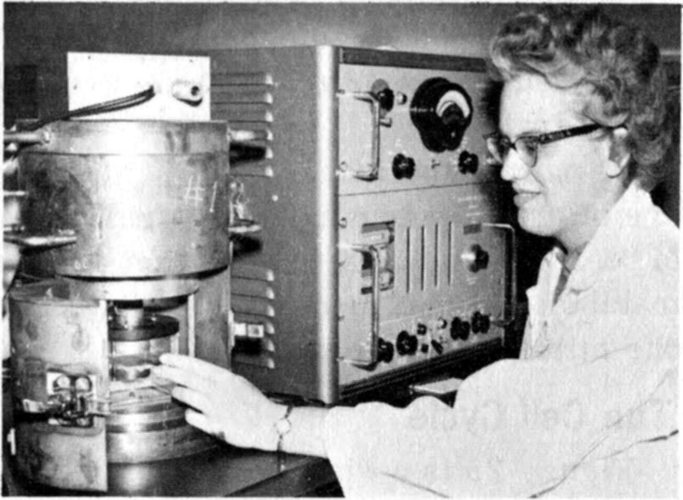
Figure 24 A college chemistry major analyzing a sample of radioactive materials with an instrument known as a proportional beta counter.
Some circumstantial evidence is very strong, as when you find a trout in the milk.
Henry David Thoreau
For a biologist interested in the mechanism of cell proliferation, the most important event in the life of a cell was, until very recently, cell division. As we mentioned, when a cell divides into two daughter cells, it undergoes a process called mitosis; mitosis itself is subdivided into four stages called prophase, metaphase, anaphase, and telophase. Mitosis in most cells takes less than one hour. Between one mitosis and the next, there can be an interval, from a few hours to several days in length, during which a cell is said to be in interphase. The entire period between the midpoints of two successive mitoses is called the cell cycle.
Until a few years ago, we knew very little about interphase. In fact, in one classic book on histology,[8] while a description of mitosis required almost 12 pages, interphase was dismissed in less than six lines! The reason for this lack of interest was, of course, the fact that no adequate methods were available for studying metabolic activities of cells in interphase. The methods of high-resolution autoradiography and radiochemical analysis of synchronized cell populations have become available only in the past few years.
We now know that metabolic activities during interphase are of primary importance in understanding the mechanism of cell division. It is, in fact, the orderly sequence of metabolic events occurring in interphase that leads from one mitosis to the next.
Figure 25 is a diagram of the cell cycle. Try to imagine the cell cycle as a race track and individual cells as cars that race around it. You are sitting at the finish wire, which is mitosis (we chose mitosis because it is easy to recognize when the cell is observed with the aid of a microscope). At a certain time during the race, all the cars in a portion of the track, say a 200-yard sector of the backstretch, are sprayed with a blue dye as they race by. These cars are now marked, just as cells synthesizing DNA are marked if briefly exposed to tritiated thymidine, the common radioactive precursor of DNA. As soon as these cars have been sprayed, you observe all the cars as they pass the finish line in front of you. At first, you will see cars that were nearest the wire and were not sprayed; then the dye-marked cars will pass; and finally more unmarked cars, those that had passed the finish line but had not reached the spray area when the marking was done, will come by. If you replace the words spray, cars, and wire with the words radioactivity, cells, and mitosis, you have described the cell cycle and the flow of cells in the cycle.
Now, if all cars were going at the same speed, you could calculate with great accuracy the time taken for any one car to go around the track, or from the finish line to the backstretch, or through the spray sector, and so on. However, since cars move at different speeds, you can only obtain an average time for all sprayed cars. Similarly, since individual cells behave differently, you can only obtain averages of the times these cells spend in the various portions of the cell cycle.
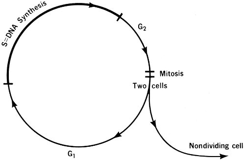
Figure 25
THE CELL CYCLE
These cell-cycle portions are four in number, according to nomenclature originated by A. Howard and S. R. Pelc, two English investigators who first described the cycle: (1) mitosis; (2) G₁, which is the period between mitosis and DNA synthesis; (3) S phase, which is the period during which DNA is replicated; and (4) G₂, which is the period between DNA synthesis and the next mitosis. Only cells in the S phase (DNA synthesis) are marked when exposed to a radioactive precursor of DNA.
Because it has several important implications in biology and medicine, it is important to remember that DNA synthesis occurs only during the short, well-defined S period of the cell cycle. Other synthetic processes go on 40 throughout the cycle. We mentioned, for instance, that all cells can be labeled by a brief exposure to a radioactive amino acid, a precursor of proteins; this means that protein synthesis occurs throughout the entire cell cycle, including mitosis. When we use a radioactive RNA precursor, all cells except those in anaphase and metaphase are labeled; this means that RNA synthesis occurs throughout the entire cycle except during anaphase and metaphase. But a radioactive tag on a DNA precursor reveals that only during the S phase is there DNA synthesis.[9]
It is also important to remember that a cell that has synthesized DNA is a cell that, with a few exceptions, will divide in the very near future. Thus, for an understanding of the mechanisms that control cellular proliferation, it is important to investigate the factors that control DNA synthesis. Our recent knowledge of the cell cycle has therefore led to a shift in the focus of investigation from mitosis to DNA synthesis.
Another point to remember is that not all cells keep going through the cell cycle indefinitely. As shown in Figure 25, when a cell divides, the daughter cells have two alternatives, either to go through another cycle or to leave it altogether. Cells that leave the cycle are called differentiated cells and will eventually die without any further division. Many cells in an adult organism also have lost the capacity to make DNA and therefore the capacity to divide. These cells often have other specialized functions in the body; examples are nerve cells and muscle cells.
The synthesis of other macromolecules (giant molecules, like DNA) connected with the gene-action system is another field of active investigation. We have described how we can investigate the synthesis of proteins and RNA with radioactive isotopes, and we have given some information on the gene-action system, which is also shown in Figure 26.
The genetic material of a cell is DNA. The DNA molecule is in the form of a double-stranded helix that is supported by a protein backbone. Genes are often described 41 as simply segments of DNA. They differ from each other only in the order in which the four nucleotide bases that make up DNA are arranged. (Look at Figure 13 again.) Since a single gene is usually made up of several hundred bases, it is easy to imagine the infinite variety of genes that could exist by simply changing the order of the four bases several hundred times.
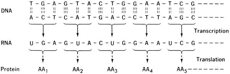
Figure 26
THE GENE-ACTION SYSTEM
Not all genes in the cells of a living organism are active. In fact, most of them are inactive, or, as geneticists say, repressed. What represses genes to make them inactive is not known, but many investigators believe the activity, or lack of it, is regulated by proteins called histones. If a gene is repressed, nothing happens; it remains inactive, presumably until something removes the repressing factor. But an active gene sets in motion a train of events that results in activation of one of the processes of life: The gene’s DNA directs the manufacture of RNA, which in turn brings about the synthesis of a specific protein to carry out a specific metabolic process. In other words, all the activities of the cell are dictated by active genes (the DNA molecules) through the mediation of RNA and are executed by proteins.
Here is what happens as nearly as scientists can reconstruct it:
The DNA of a particular active gene manufactures a molecule of m-RNA by the same kind of replication that it uses for making more DNA. In m-RNA the sequence of bases is the same as in the parent DNA segment; for this 42 reason, m-RNA is also called DNA-like RNA. As shown in Figure 12, a cytosine molecule in m-RNA corresponds to a cytosine molecule in DNA, a guanine to a guanine, and so on, except that the m-RNA has uracil in all the places where thymine occurs in DNA. The order of the nucleotides in the m-RNA is the same as that in the DNA, so the m-RNA carries the genetic code of the gene that made it. This process, all of which occurs in the cell nucleus, is one of copying, or transcription, rather than translation, since the same “codewords” (the nucleic-acid bases) are reproduced.
The new m-RNA molecule then travels from the nucleus to the cytoplasm and attaches itself to an unoccupied ribosome (see Figure 27). Here it fits to a molecule of r-RNA and blends its shape geometrically, or spatially, with the shape of the r-RNA in lock-and-key, or jigsaw-puzzle, fashion. The combined new RNA molecule is now capable of manufacturing a specific protein.
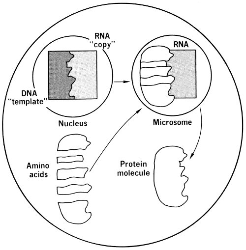
Figure 27 Protein synthesis in a ribosome (microsome), and its control by DNA in the nucleus, using RNA as an intermediary.
Adapted from Principles of Biology, Neal D. Buffaloe, Prentice-Hall, Inc., 1962, with permission.
At this point an s-RNA molecule arrives, bringing with it one amino-acid molecule, which then combines with other 43 amino acids in the specific order dictated by the RNA to form a specific protein. After the amino acids have been formed into the protein molecule, they detach themselves from the s-RNA molecule. The s-RNA molecule has two recognition sites by which it matches up to its neighbors: One recognizes, or “fits”, the amino acid, and the other recognizes a corresponding triplet of bases on m-RNA. There is thus a particular s-RNA molecule for each amino acid and a particular triplet of bases on the m-RNA molecule for each triplet of bases that is specific to the s-RNA molecule.
In this process the machinery has translated the nucleic-acid code into the protein code; that is, it has translated a sequence of the bases into a sequence of amino acids. This process is therefore called translation of the genetic message. Once the protein has been synthesized, it will become active in performing some of the cell’s metabolic activities.
The gene-action system actually is somewhat more elaborate than this. There are feedback mechanisms, genes that control the activity of other genes, either directly or through the production of specific proteins, and so on. However, the scheme just outlined gives a fair, if simplified, idea of how the genetic message is carried to the entire cell and how it is translated into actual life processes.
... a riddle wrapped in a mystery inside an enigma.
Winston Churchill
The various procedures in which radioactive isotopes play a major role have been applied to many studies and investigations in the fields of biology and medicine. In fact, most of the concepts of modern biology that we have been discussing in this booklet owe their discovery to the judicious use of radioisotopes. To illustrate how radioisotopes can be used to solve a practical problem, we 44 have chosen a typical example, the investigation, at a molecular level, of the effectiveness of an anti-cancer drug.
Several drugs that exert a beneficial effect, at least temporarily, on the course of certain cancers have been used by doctors for several years. Most of them were discovered empirically, that is, by accident, during routine trials against cancers. Doctors know they work but do not always know how. They would also like to know the mechanism of the drugs’ action at the molecular level so that the knowledge might open the way to the discovery of other drugs more effective against cancer and less toxic against normal cells. The following experiment shows how the molecular effect of an anti-cancer drug is studied.
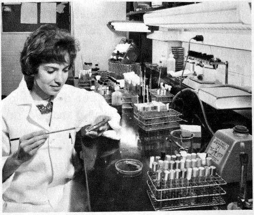
Figure 28 Technician preparing tissues for comparative studies.
Cells growing in tissue cultures are often used to test anti-cancer drugs (see Figure 28). These cells, derived from human cell lines, are grown in glass or plastic bottles as a suspension in a nutrient medium. To begin, a culture is divided into halves. To one half is added the anti-cancer drug Actinomycin D. The other half will continue to grow without addition of other substances and will serve as a control, or comparison. After a suitable time has elapsed for the drug to act on the cultured cells, similar portions of the drug-treated cells and the control cells will be tested in several ways. One portion of each kind of cells is incubated with ³H-thymidine to determine the effect of the drug on DNA synthesis. Two other portions 45 are incubated with ³H-cytidine to study the effect on RNA synthesis. Another pair will be tested with ¹⁴C-leucine to investigate protein synthesis. The effect of the drug, of course, is determined by comparing the untreated control with the drug-treated culture.
The biochemical, autoradiographic, and counting techniques that we described previously are all used to determine the uptake of the radioisotopes into the cell’s components. Chromatography is used to ascertain if the drug has changed the concentration of precursors (thymidine, cytidine, or leucine) in the nutrient medium, since a change in these could produce misleading results. Finally, if the drug is found to have an effect on RNA, we can investigate the type of RNA that is affected by centrifuging phenol-purified RNA.
The results will disclose the primary site (DNA, RNA, or proteins) of the drug action on cell metabolism. More elaborate experiments can pinpoint more intimately the mechanism of action. By studying the life processes of cells, we can advance toward a common denominator in anti-cancer drugs that will lead to an effective anti-cancer treatment.
Thus, the task is, not so much to see what no one has seen yet; but to think what nobody has thought yet, about what everybody sees.
Arthur Schopenhauer
The use of radioactive isotopes in the study of life processes is of importance in understanding them. With the use of autoradiographic and radiochemical techniques, it is possible to obtain valuable information regarding the life of cells and the intimate mechanisms by which life processes determine the fate of the entire organism.
Our knowledge of the cell cycle and of the gene-action system has been useful in determining how organisms grow and how cancer cells behave. It has been determined that certain normal adult cells divide more frequently 46 than some cancer cells and that the growth of cancers depends not so much on the speed of cellular proliferation as on the number of cells actually dividing.
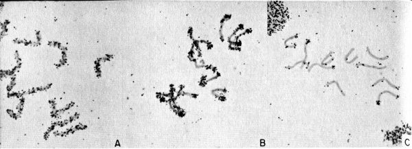
Figure 29 Radioautograph showing DNA synthesis during chromosome replication. Chromosomes from cells in the root tip of the Tradescantia plant were labeled with ³H-thymidine. In A and B, the midportion of DNA synthesis, the radioisotope is distributed throughout the chromosome arms; in C, near the end of DNA synthesis, it is confined mainly to the end of the arms.
Knowledge of the cell cycle has also brought new insight to the control of cell division, as in studies related to the therapy of cancer. The most important problem now is, not the control of cell division, but the control of the synthesis of DNA.
Our information on the gene-action system provides broad new opportunity for the investigation of many life processes. Hormone action, processes by which the body develops immunity to disease, and even cell division itself are apparently regulated through the gene-action system. This, in turn, offers possibilities for investigations meant to control these processes.
It is difficult to chart the future course of modern molecular biology, but it is not difficult to predict that the next few years will bring to biology the same kind of sweeping advances that revolutionized physics a few decades ago. The DNA molecule has been called the atom of life. When we have harnessed it, the harnessing of the uranium atom will seem, in comparison, a result of scientific adolescence. When man has mastered the genetic code, he’ll hold a vast power in his hands—power over the nature of coming generations.
The Cell, Carl P. Swanson, Prentice-Hall, Inc., Englewood Cliffs, New Jersey, 1964, 114 pp., $1.75.
Inside the Living Cell, J. A. V. Butler, Basic Books, Inc., New York, 1959, 174 pp., $3.95.
Life and Energy, Isaac Asimov, Doubleday & Company, Inc., Garden City, New York, 1962, 380 pp., $4.95.
Applied Nuclear Physics, Ernest C. Pollard and William L. Davidson, John Wiley & Sons, Inc., New York, 1956, 352 pp., $6.00.
Adventures in Radioisotope Research, the collected works, with recent annotations, of George de Hevesy, Pergamon Press, Inc., New York, 1961, 1047 pp. (2 volumes), $30.00.
The Biochemistry of Nucleic Acids, J. N. Davidson, John Wiley & Sons, Inc., New York, 4th edition, 1960, 287 pp., $4.25.
The Machinery of the Body, A. J. Carlson and C. Johnson, The University of Chicago Press, Chicago, Illinois, 1961, 752 pp., $6.50.
Life: An Introduction to Biology, George G. Simpson and William S. Beck, Harcourt, Brace & World, Inc., New York, 2nd edition, 1965, 869 pp., $8.95.
From Cell to Test Tube, Robert W. Chambers and Alma Payne, Charles Scribner’s Sons, New York, 1962, 216 pp., $1.45.
Isotopic Tracers in Biology, M. D. Kamen, Academic Press Inc., New York, 3rd edition, 1957, 474 pp., $9.50.
Autoradiography in Biology and Medicine, G. A. Boyd, Academic Press Inc., New York, 1955, 399 pp., $10.00.
A Tracer Experiment: Tracing Biochemical Reactions with Radioisotopes, Martin D. Kamen, Holt, Rinehart & Winston, Inc., New York, 1964, 127 pp., $1.28.
Molecular Biology: Genes and the Chemical Control of Living Cells, J. M. Barry, Prentice-Hall, Inc., Englewood Cliffs, New Jersey, 1964, 139 pp., $3.35.
Elementary Biophysics: Selected Topics, Herman T. Epstein, Addison-Wesley Publishing Company, Inc., Reading, Massachusetts, 1963, 122 pp., $2.95 (hardback), $1.75 (paperback).
Autobiographies of Cells, R. Baserga and W. Kisieleski, Scientific American, 209: 103 (August 1963).
Electrons, Enzymes, and Energy, Michael G. Del Duca and John M. Fuscoe, International Science and Technology, 39: 56 (March 1965).
Scientific American, 205 (September 1961). This is a special issue on the living cell. The two articles cited below are of particular interest:
Liquid Scintillation Counting: Proceedings of a Conference Held at Northwestern University, August 20-22, 1957, C. G. Bell, Jr. and F. N. Hayes (Eds.), Pergamon Press, Inc., New York, 1957, 292 pp., $10.00.
Atomic Energy Research: Life and Physical Sciences; Reactor Development; and Waste Management, A Special Report of the U. S. Atomic Energy Commission (December 1961), Superintendent of Documents, U. S. Government Printing Office, Washington, D. C. 20402, 333 pp., $2.25.
Radioisotopes in the Service of Man, Fernand Lot, National Agency for International Publications, 317 East 34th Street, New York 10016, 1958, 82 pp., $1.00.
Science and Cancer, M. B. Shimkin, Public Health Service Publication No. 1162, Superintendent of Documents, U. S. Government Printing Office, Washington, D. C. 20402, 1964, 137 pp., $0.60.
The Cell: Structural Unit of Life, 10 minutes, sound, color or black and white, 1949, Coronet Films, Inc., 65 E. South Water Street, Chicago, Illinois 60601.
Continuity of Life: Characteristics of Plants and Animals, 11 minutes, sound, color or black and white, 1954, Audio-Visual Center, Indiana University, Bloomington, Indiana 47405.
DNA: Molecule of Heredity, 16 minutes, sound, color (No. 1825), black and white (No. 1826). 1960, Encyclopaedia Britannica Films, Inc., Wilmette, Illinois 60091.
The Science of Genetics, AIBS Secondary School Film Series, No. 13280, 25 minutes, sound, color, 1962, McGraw-Hill Book Company, Inc., 330 West 42nd Street, New York 10036.
Available for loan without charge from the AEC Headquarters Film Library, Division of Public Information, U. S. Atomic Energy Commission, Washington, D. C. 20545, and other AEC film libraries:
Tracing Living Cells, Challenge Film No. 11, 29 minutes, sound, black and white, 1962. Produced by Ross-McElroy Productions 49 for the National Educational Television and Radio Center under a grant from Argonne National Laboratory. This nontechnical film demonstrates some of the uses of radioisotopes in the study of cell division and in medical therapy.
The Eternal Cycle, 12½ minutes, sound, black and white, 1954. Produced by the Handel Film Corporation. This nontechnical film illustrates the use of radioisotope tracers in biological research and is suitable for intermediate- through college-level audiences.
Chromosome Labeling by Tritium, 15 minutes, sound, color, 1958. Produced by the Jam Handy Organization for the U. S. Atomic Energy Commission. This technical film discusses the advantages of tritium over other radioisotopes as labeling material in autoradiography.
A is for Atom, 15 minutes, sound, color, 1953. Produced by the General Electric Company. This nontechnical film explains the structure of the atom, natural and artificially produced elements, stable and unstable atoms, principles and applications of nuclear reactors, and the benefits of atomic radiation to biology, medicine, industry, and agriculture. It is suitable for elementary- through high-school audiences.
This booklet is one of the “Understanding the Atom” Series. Comments are invited on this booklet and others in the series; please send them to the Division of Technical Information, U. S. Atomic Energy Commission, Washington, D. C. 20545.
Published as part of the AEC’s educational assistance program, the series includes these titles:
A single copy of any one booklet, or of no more than three different booklets, may be obtained free by writing to:
USAEC, P. O. BOX 62, OAK RIDGE, TENNESSEE 37830
Complete sets of the series are available to school and public librarians, and to teachers who can make them available for reference or for use by groups. Requests should be made on school or library letterheads and indicate the proposed use.
Students and teachers who need other material on specific aspects of nuclear science, or references to other reading material, may also write to the Oak Ridge address. Requests should state the topic of interest exactly, and the use intended.
In all requests, include “Zip Code” in return address.
Printed in the United Slates of America
USAEC Division of Technical Information Extension, Oak Ridge, Tennessee