
BOOKS ON POPULAR SCIENCE
By CHARLES R. GIBSON, F.R.S.E.
“Mr. Gibson has a fine gift of exposition.”—Birmingham Post.
“Mr. Gibson has fairly made his mark as a populariser of scientific knowledge.”—Guardian.
In the Science for Children Series. Illustrated. 4s. 6d. nett each.
OUR GOOD SLAVE ELECTRICITY.
THE GREAT BALL ON WHICH WE LIVE.
THE STARS & THEIR MYSTERIES.
WAR INVENTIONS & HOW THEY WERE INVENTED.
CHEMISTRY & ITS MYSTERIES.
In the Science of To-Day Series. Illustrated. 7s. 6d. nett each.
SCIENTIFIC IDEAS OF TO-DAY. A Popular Account of the Nature of Matter, Electricity, Light, Heat, &c. &c.
“Explained without big words, with many interesting pictures, and with an ease in exposition.”—Scotsman.
ELECTRICITY OF TO-DAY; Its Work and Mysteries described in Non-technical Language. With 30 Illustrations.
“A masterly work.”—Globe.
In the Romance Library. Illustrated. 6s. nett each.
THE ROMANCE OF MODERN ELECTRICITY. What is known about Electricity and many of its interesting applications.
“Clear and concise.”—Graphic.
THE ROMANCE OF MODERN PHOTOGRAPHY.
“There is not a dry or uninteresting page throughout.”—Country Life.
“The narration is everywhere remarkable for its fluency and clear style.”—Bystander.
THE ROMANCE OF MODERN MANUFACTURE.
“A popular and practical account of all kinds of manufacture.”—Scotsman.
THE ROMANCE OF SCIENTIFIC DISCOVERY.
HEROES OF THE SCIENTIFIC WORLD. The Lives, Sacrifices, Successes, and Failures of some of the greatest Scientists. With 19 Illustrations.
“The whole field of science is well covered.... Every one of the 300 and odd pages contains some interesting piece of information.”—Athenæum.
WHAT IS ELECTRICITY. Long 8vo. With 8 Illustrations. 6s. nett.
“A brilliant study.”—Daily Mail.
“Quite a unique book in its way, at once attractive and illuminating.”—Record.
THE MARVELS OF PHOTOGRAPHY. Illustrated. 5s. nett.
In the Wonder Library. Illustrated. 3s. nett each.
THE WONDERS OF MODERN MANUFACTURE.
THE WONDERS OF WAR INVENTIONS.
THE WONDERS OF MODERN ELECTRICITY. With 17 Illustrations and Diagrams.
WIRELESS TELEGRAPHY. A Popular Description of Wireless Telegraphy and Telephony in which no technical terms are used, and no previous knowledge of the subject assumed. 3s. 6d. nett.
THE SCIENCE OF TO-DAY SERIES
With many Illustrations. Extra Crown 8vo. 7s. 6d. nett
New Volume
SUBMARINE WARFARE OF TO-DAY. Telling how the Submarine Menace was met & vanquished. By C. W. Domville-Fife, Staff of H.M. School of Submarine Mining. With 53 Illustrations.
“A very striking book, revelation follows revelation, and magnificent stories of fighting & heroism at sea come practically on every page. One of the few war books which will survive the next 10 years.”—Liverpool Courier.
AIRCRAFT OF TO-DAY. A Popular Account of the Conquest of the Air. By Maj. Charles C. Turner, R.A.F. With 62 Illustrations.
“Maj. Turner is well known as an authority on aeronautics. Of real value.”—Aberdeen Journal.
GEOLOGY OF TO-DAY. A Popular Introduction in Simple Language. By J. W. Gregory, F.R.S., D.Sc., Professor of Geology at the University of Glasgow. With 55 Illustrations. Extra Crown 8vo.
“An ideal introduction to a fascinating science. The romance and reality of the earth most brilliantly and soundly presented.”—Globe.
SUBMARINE ENGINEERING OF TO-DAY. By C. W. Domville-Fife, Author of “Submarines of the World’s Navies,” &c.
BOTANY OF TO-DAY. A Popular Account of the Evolution of Modern Botany. By Prof. G. F. Scott-Elliot, M.A., B.Sc., F.L.S.
“This most entertaining and instructive book. It is the fruit of wide reading and much patient industry.”—Globe.
SCIENTIFIC IDEAS OF TO-DAY. A Popular Account, in Non-technical Language, of the Nature of Matter, Electricity, Light, Heat, Electrons, &c. &c. By C. R. Gibson, F.R.S.E. Extra Crown 8vo.
“As a knowledgeable writer, gifted with the power of imparting what he knows in a manner intelligible to all, Mr. C. R. Gibson has established a well-deserved reputation.”—Field.
ASTRONOMY OF TO-DAY. A Popular Introduction in Non-technical Language. By Cecil G. Dolmage, LL.D., F.R.A.S. 46 Illustrations. Extra Crown 8vo.
“A lucid exposition much helped by abundant illustrations.”—The Times.
ELECTRICITY OF TO-DAY. Its Work and Mysteries Explained. By Charles R. Gibson, F.R.S.E. Extra Crown 8vo.
“One of the best examples of popular scientific exposition that we remember seeing.”—The Tribune.
ENGINEERING OF TO-DAY. A Popular Account of the Present State of the Science. By T. W. Corbin. 39 Illustrations. Ex. Cr. 8vo.
“Most attractive and instructive.”—Record.
MEDICAL SCIENCE OF TO-DAY. A Popular Account of recent Developments. By Willmott-Evans, M.D., B.Sc., F.R.C.S.
“A very Golconda of gems of knowledge.”—Manchester Guardian.
MECHANICAL INVENTIONS OF TO-DAY. An Interesting Description of Modern Mechanical Inventions. By Thomas W. Corbin.
“In knowledge and clearness of exposition it is far better than most works of a similar character and aim.”—Academy.
PHOTOGRAPHY OF TO-DAY. A Popular Account of the Origin, Progress, and Latest Discoveries. By H. Chapman Jones, F.I.C., F.C.S., Pres. R.P.S.; Lecturer on Photography at Imperial College of Science.
“An admirable statement of the development of photography from its very beginning to the present time.”—Journal of Photography.
SEELEY, SERVICE & CO., LIMITED
THE ROMANCE OF THE
MICROSCOPE
By the courtesy of Messrs. F. Davidson & Co.
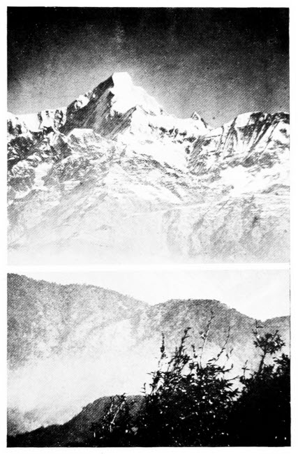
An Example of a Micro-Telescopic Photograph
“Nanda Kot.” Height, 22,510 feet; distance, 60 miles. The trees at the lower right-hand corner are only 20 yards from the photographer. A remarkable photograph, showing the great depth of focus of the micro-telescope.
AN INTERESTING DESCRIPTION OF ITS
USES IN ALL BRANCHES OF SCIENCE,
INDUSTRY, AGRICULTURE, AND IN THE
DETECTION OF CRIME, WITH A
SHORT ACCOUNT OF ITS ORIGIN,
HISTORY & DEVELOPMENT.
BY
C. A. EALAND, M.A.
AUTHOR OF “ANIMAL INGENUITY OF TO-DAY,”
“INSECTS AND MAN,” &C., &C.
WITH 39 ILLUSTRATIONS & NUMEROUS DIAGRAMS
LONDON
SEELEY, SERVICE & CO. LIMITED
38 GREAT RUSSELL STREET
1921
UNIFORM WITH THIS VOLUME
THE LIBRARY OF ROMANCE
Extra Crown 8vo. With many illustrations. 6s. nett.
“Splendid Volumes.”—The Outlook.
“The Library of Romance offers a splendid choice.”—Globe.
“Gift Books whose value it would be difficult to over-estimate.”—The Standard.
“This series has now won a considerable & well deserved reputation.”—The Guardian.
“Each Volume treats its allotted theme with accuracy, but at the same time with a charm that will commend itself to readers of all ages. The root idea is excellent, and it is excellently carried out, with full illustrations and very prettily designed covers.”—The Daily Telegraph.
The Romance of Savage Life
The Romance of Plant Life
The Romance of Early British Life
The Romance of Modern Sieges
The Romance of Bird Life
The Romance of Animal Arts and Crafts
The Romance of the World’s Fisheries
The Romance of Missionary Heroism
The Romance of Polar Exploration
The Romance of Modern Photography
The Romance of Modern Electricity
The Romance of Modern Manufacture
The Romance of Scientific Discovery
The Romance of Aeronautics
The Romance of Modern Astronomy
The Romance of the Post Office.
The Romance of Early Exploration
The Romance of Modern Exploration
The Romance of Modern Mechanism
The Romance of Modern Invention
The Romance of Modern Engineering
The Romance of Modern Locomotion
The Romance of Modern Mining
The Romance of the Animal World
The Romance of Insect Life
The Romance of the Mighty Deep
The Romance of Modern Geology
The Romance of Modern Chemistry
The Romance of the Ship
The Romance of Piracy
The Romance of Submarine Engineering
The Romance of War Inventions
The Romance of the Spanish Main
The Romance of Modern Commerce.
SEELEY, SERVICE & CO., LIMITED.
| CHAPTER I | PAGE |
| Early Days of the Microscope | 17 |
| CHAPTER II | |
| Some Early Microscopists | 29 |
| CHAPTER III | |
| The Action of Light | 40 |
| CHAPTER IV | |
| The Compound Microscope | 50 |
| CHAPTER V | |
| Animal Life in Ponds and Streams | 66 |
| CHAPTER VI | |
| Plant Life in Ponds and Streams | 83 |
| CHAPTER VII | |
| The Microscope and Plant Life | 97 |
| CHAPTER VIII | |
| [10]Animal Life and the Microscope | 112 |
| CHAPTER IX | |
| The Study of the Rocks | 125 |
| CHAPTER X | |
| The Microscope as Detective | 137 |
| CHAPTER XI | |
| Bacteria | 152 |
| CHAPTER XII | |
| Medical Work with the Microscope | 167 |
| CHAPTER XIII | |
| The Microscope and Agriculture | 178 |
| CHAPTER XIV | |
| The Microscope and Insect Life | 192 |
| CHAPTER XV | |
| The Microscope by the Seaside—Animal Life | 208 |
| CHAPTER XVI | |
| The Microscope by the Seaside—Plant Life | 225 |
| CHAPTER XVII | |
| Micro-Telescope and Super Microscope | 239 |
| CHAPTER XVIII | |
| [11]Chemistry and the Microscope | 248 |
| CHAPTER XIX | |
| Use of the Microscope in Manufactures | 260 |
| CHAPTER XX | |
| The Microscope and Camera allied | 274 |
| CHAPTER XXI | |
| How the Glass used in Microscopes is made | 282 |
| CHAPTER XXII | |
| The Choice and Use of Apparatus | 291 |
| PAGE | |
| Nanda Kot—An Example of Tele-Photography | Frontispiece |
| Head of Dog Flea | 56 |
| Starch Grains of Potato | 72 |
| Phosphorescence Animalculæ | 72 |
| Proteus Animalcule | 72 |
| Cyclops | 72 |
| Bladderwort | 88 |
| Spores of Horse-tail | 88 |
| Hairs on a Potato Leaf | 88 |
| Spirogyra | 88 |
| Thorn Insect | 120 |
| Head of Palm Weevil | 120 |
| [14]Leaf Insect | 120 |
| Head of Stick Insect | 120 |
| Foraminifera | 128 |
| Diatoms | 128 |
| Stinging Hairs of Nettle | 144 |
| Butterfly Wing Scales | 144 |
| Crystals from Human Blood | 168 |
| Crystals from the Blood of the Baboon | 168 |
| Cluster Cups | 184 |
| Rust of Wheat | 184 |
| Pollen Grains on a Grass Flower | 184 |
| Lower Side of a Fern Frond | 184 |
| Head of a Beetle | 200 |
| Head of Hercules Beetle | 200 |
| A Cicada | 200 |
| Head of Mantis | 200 |
| Face of a Fly | 216 |
| [15]Section of Human Skin | 216 |
| Feeler of Cockchafer | 232 |
| View with Ordinary Camera | 240 |
| View with Micro-Telescope | 240 |
| Eye of a Cockchafer | 256 |
| Hooks on a Bee’s Wing | 256 |
| Spider’s Foot | 264 |
| Fly’s Foot | 264 |
| Fly’s Eye | 296 |
| Images seen by a Fly | 296 |
The Romance of the Microscope
It is certain that lenses were used as early as the thirteenth century, and it is probable that they date back to far earlier times. The ancient gem cutters probably used spheres of glass filled with water as magnifiers, their work could hardly have been accomplished without some artificial aid. We know, from early writings, that burning glasses were used by physicians in their work, and Seneca, the author, who wrote in A.D. 63, says: “Letters, however small and dim, are comparatively large and distinct when seen through a glass globe filled with water.”
Euclid, whose name at least is familiar to everyone, was, as shown by his writings, perfectly well acquainted with the fact that curved mirrors may be used to magnify objects, and that was so long ago as the third century B.C. Convex glasses, used as spectacles, were first mentioned by Bernard de Gordon, about 1307, but, as far as we know, they[18] were never used for the purpose of studying minute living objects.
To Leonardo da Vinci belongs the honour of seriously investigating, for the first time, the properties of concave and convex lenses, and several alchemists, as the early chemists were called, used flasks filled with water, concave mirrors or glass balls to gather together the rays of the sun. “Long before the dawn of the seventeenth century, the principle of the lens was both comprehended and applied to scientific matters by the Englishmen, Leonard Digges and his son Thomas, and by the Italian, Giambattista Porta.”
Towards the end of the sixteenth century and the early part of the seventeenth century, interest in the minute structure of natural objects appears to have developed. As early as 1590, Thomas Mouffet used magnifying glasses in studying small mites, and in 1637 Descartes invented a single lens microscope in which the rays of light were reflected on to the object by means of a concave mirror. This method of illumination, it is interesting to note, is still used in some forms of pocket magnifiers. Most of the early discoveries were made with single lenses, for in the compound microscopes which were first made, it was only possible to view such a small portion of an object at one time that the advantage lay with the less complicated instrument.
The earliest microscopes were simply short tubes of any material which would not admit light; at one end there was a lens, at the other a glass plate on[19] which the object to be examined was placed. Because these crude instruments were chiefly used for the examination of insects they were known as “Vitrea pulicaria” or “Vitrea muscaria.” Later they were called “Engyoscopes,” and, after the invention of compound microscopes, they were described as “Microscopia ludicra,” as opposed to the latter instruments, known as “Microscopia seria.”
The next stage in the development of the microscope consisted in the introduction of lenses of very short focal length, and, in 1665, Robert Hooke used small glass balls, formed by fusing threads of drawn glass, for this purpose.
It was Antony van Leeuwenhoek, however, who perfected these instruments. He brought an extraordinary skill and industry to bear on the grinding and polishing of minute lenses of short focal length. Already in 1673 Regnier de Graaf wrote to the Royal Society in London that Leeuwenhoek was making glasses far superior to those of the great Italian lens maker, Eustachio Divini. Leeuwenhoek’s success was largely due not only to his method of grinding, but also to the skill with which he mounted his lenses, which were accurately fitted into a minute hole in a metal plate. The object to be examined was firmly held in a stand and adjusted by means of a screw movement. By this means, and by the use of hollow metal reflectors, he succeeded in availing himself of transmitted light in the case of transparent objects. Leeuwenhoek was able to make immense advances with these instruments,[20] the minute pond animals he could see with ease, and by 1683 he had even attained a sight of the bacteria. His researches represented the high-water mark of work done with the simple microscope, most of the later work was carried out with the compound instrument.
The earliest history of the compound microscope is difficult to separate from that of the telescope and, in any complete account, the two instruments must be considered together. It appears that the first scientist that conceived the idea of using a series of lenses, rather than a single lens, was Leonard Digges, whom we have already mentioned.
In a book by Porta, a writer who though not himself original, was gifted with great curiosity and industry in the collection of the ideas of others, we read: “How to make plain a letter held far away by means of a lens of crystal,” and also that “with a concave lens you see things afar smaller but plainer, with a convex lens you see them larger but less distinct. If, however, you know how to combine the two sorts properly you will see near and far both large and clear.”
Shortly after the publication of Porta’s book the method of combining two lenses into a microscope or telescope was discovered, quite accidentally, by a Dutch boy named Zacharias, who worked in the shop of his father, a spectacle maker. The event was described by Willem Boreel, Dutch Ambassador to France, in a letter written in 1655. He wrote: “I am a native of Middleburg, the capital of[21] Zeeland, and close to the house where I was born, there lived in the year 1591 a certain spectacle maker, Hans by name. His wife, Maria, had a son, Zacharias, whom I knew very well, because I constantly as a neighbour and from a tender age went in and out playing with him. This Hans or Johannes with his son Zacharias, as I have often heard, were the first to invent microscopes, which they presented to Prince Maurice, the governor and supreme commander of the United Dutch forces, and were rewarded with some honorarium. Similarly they afterwards offered a microscope to the Austrian Archduke Albert, supreme governor of Holland. When I was Ambassador to England in the year 1619, the Dutchman Cornelius Drebbel of Alkomar, a man familiar with many secrets of nature, who was serving there as a mathematician to King James, and was well known to me, showed me that very instrument which the Archduke had presented as a gift to Drebbel, namely, the microscope of Zacharias himself. Nor was it (as they are most seen) with a short tube, but nearly two and a half feet long, and the tube was of gilded brass two fingers’ breadth in diameter, and supported on three dolphins formed also of brass. At its base was an ebony disc, containing shreds or some minute objects which we inspected from above, and their forms were so magnified as to seem almost miraculous.” So this was the first compound microscope!
Although Zacharias invented the microscope, it was Galileo who introduced it to the scientific world.[22] He published a book in 1610 in which he wrote: “About ten months ago a rumour reached me of an ocular instrument made by a certain Dutchman, by means of which an object could be made to appear distinct and near to an eye that looked through it, although it was really far away. And so I considered the desirability of investigating the method, and reflected on the means by which I might come to the invention of a similar instrument. I first prepared a leather tube at the ends of which one placed two lenses each of them flat on one side, and as to the other side I fashioned one concave and the other convex. Then holding the eye to the concave one, I saw the objects fairly large and nearer, for they appeared three times nearer and nine times larger than when they were observed by the naked eye. Soon after I made another more exactly, representing objects more than sixty times larger. At length, sparing no labour and no expense, I got to the point that I could construct an excellent instrument so that things seen through it appeared a thousand times greater and more than thirty-fold nearer than if observed by the naked eye.” Galileo had his enemies, who accused him of having picked Zacharias’s brains; he admitted that he had taken his idea from the Dutchman’s invention, but further than that he would not go; in fact, he replied that the invention of Zacharias was a mere accident but that his own instrument was discovered by a process of reasoning.
It would serve no good purpose to tell the story[23] of all the scientists who have helped to bring the microscope to its present state of perfection, although many of their descriptions of objects and apparatus are as quaint as the latter. Scheiner, for example, who wrote in 1630, mentions “that wonderful instrument the microscope, by means of which a fly is magnified into an elephant, and a flea into a camel.” To Kircher belongs the credit of being the first worker to construct an instrument with coarse and fine adjustment and with a substage condenser, which could be used either for concentrating the sun’s rays or those from a lamp. With an instrument of this pattern Malpighi saw the circulation of blood in a frog’s lung. By 1685, when instruments with four and six lenses were being used, the compound microscope was firmly established as a help to scientists, and the simple lens was used thereafter as an adjunct but not a rival to the newer instrument.
History makes a strong appeal to many people, and those who are fascinated thereby will find endless amusement in reading old books on the microscope and its objects. In the preface to Mouffet’s Insectorum Theatrum, one of the earliest books on insects, we read the following quaint lines: “If you will take lenticular object glasses of Crystal (for though you have Lynx his eyes, they are necessary in searching for atoms) you will admire to see the Fleas that are curasheers, and their hollow trunk to torture men, which is a bitter plague to maids, you shall see the eyes of Lice sticking[24] forth, and their horns, their bodies crammed all over, their whole substance diaphanous, and through that, the motion of their heart and blood. Also little Handworms, which are indivisible, they are so small, being with a needle prickt forth from their trenches near the pool of water which they have made in the skin, and being laid upon one’s nail, will discover by the sunlight their red heads and feet they creep withal.” The creatures called Handworms are itch mites, which tunnel in the human skin.
In our chapter on Nature Study and the Microscope we refer to the brown patches to be found on the backs of fern fronds; it is interesting to note that so long ago as 1646 Sir Thomas Browne had quite a good idea of their structure. Describing them, he said: “Whether these little dusty particles, upon the lower side of the leaves be seeds we have not yet been able to determine by any germination. But, by the help of magnifying glasses we find these dusty atoms to be round at first and fully representing seeds out of which at last proceed little mites, almost invisible, so that such as are old stand open, as being emptied of some bodies firstly included, which though discernible in Hartstongue, is more notoriously discoverable, in some differences, of Brake or Fern.”
Two years earlier a noted scientist, Hodierna, had made a special study of the eyes of insects and, considering the crude instruments with which he must have worked, his descriptions are wonderfully[25] accurate. Of the house fly he wrote: “The head is all eyes, prominent and without lids, lashes or brows. It is plumed with hairs like that of an ostrich and has two little pear shaped bodies hanging from the middle of the forehead. The proboscis which arises from the snout can be extended freely and stretched forth to suck up humours and can afterwards be directed back through the mouth and taken into the gullet. This instinct nature has given the creature according to its need, for it is without a neck and cannot stretch forth its head to obtain its food, as is also the case with the elephant.” The author’s knowledge of the house fly was evidently greater than his knowledge of the ostrich, for the bird has anything but a plumed head. The eye of the insect he compares to a white mulberry.
Another of these early workers, writing about the same time, gives a concise account of cheese mites, heading his description “On the creatures which arise in powdery cheese,” he wrote: “The powder examined by means of this instrument (the Compound Microscope) does not present the aspect of dirt, but teems with animalcula. It can be seen that these creatures have claws and talons and are furnished with eyes. The whole surface of their body is beautifully and distinctly coloured in such sort as I have never seen before, and which indeed, cannot be seen without wonder. They may be observed to crawl, eat and work and are equal in apparent size to a man’s nail. Their backs are all spiny and pricked out with various starlike markings[26] and surrounded by a rampart of hairs, all of such marvellous kind that you would say they are a work of art rather than of nature.”
At about this period the microscope was used for the first time for medical work and, as far as can be ascertained, Pierre Borel was the first to use it for this purpose, and he learned a great deal about the structure of flesh and the appearance of blood.
Of all the early writers on microscopy the man who spread abroad his knowledge of the instrument and its capabilities, more than anyone else, was Kircher, who died in 1680. He was an energetic writer, and wrote on a large number of subjects. His books dealt with magnetism, designs for a calculating machine, light, sound, history of plague, the philosopher’s stone, Egyptian antiquities, a history of China and a grammar. To all who read his book on the plague, it is clear that he had a good idea of infection; he was, in fact, the first writer who suspected it, though the microscope he used could not show him bacteria. In his book he wrote: “Everyone knows that decomposing bodies breed worms, but only since the wonderful discovery of the microscope has it been known that every putrid body swarms with innumerable vermicules, a statement which I should not have believed had I not tested its truth by experiments during many years.” The experiments he performed to prove his statement are so quaint that we give them in his own words.
Experiment I.—“Take a piece of meat which you[27] have exposed by night until the following dawn to the lunar moisture. Then examine it carefully with the smicroscope and you will find the contracted putridity to have been altered by the moon into innumerable wormlets of diverse size, which, however, would escape the sharpness of vision without a good smicroscope. The same is true of cheese, milk, vinegar and similar bodies of a putrifiable nature. The smicroscope, however, must be no ordinary one, but constructed with no less skill than diligence, as is mine which represents objects one thousand times greater than their true size.”
Experiment II.—“If you cut up a snake into small parts and macerate with rain water, and then expose it for several days to the sun and again bury it under the earth for a whole day and night and lastly examine the parts, separated and softened by putridity, by means of a smicroscope you will find the whole mass swarm with innumerable little multiplying serpents so that even the sharpest eyes cannot count them.”
Experiment III.—“Many authors claim that unwashed sage is injurious, but I have discovered the cause of this. For when, by means of the sun, I minutely examined the nature of the plant, I found the back of the leaves completely covered by raised work as with the figure of a spider’s web, and within the water appeared infinitesimal animalcules, which moving constantly came out of little buds or eggs.”
Experiment IV.—“If you examine a particle of rotten wood under the sun, you will see an immense[28] progeny of tiny worms, some with horns, some with wings, others with many feet. They have little black dots of eyes. What must their little livers and stomachs be like?”
In the light of modern discovery much of the writing of these early microscopists seems absurd. Kircher’s experiments, for example, prove nothing, and he is often hopelessly vague and sometimes incorrect in his statements. We must not be too critical, however, for some of this early work was excellent, the microscopes in use would not be tolerated at the present day, and without these pioneers microscopy would not have reached the stage it has. Rather than laugh at their efforts, we should marvel that they did so well.
Of the early British microscopists, Robert Hooke must not pass unnoticed. He was appointed Curator of the Royal Society two years after its formation, and the terms of his appointment were somewhat one-sided. He was required to “furnish the Society every day they meet with three or four experiments”; for this no pay was to be his till the Society accumulated sufficient funds to reward him.
Although compound microscopes had been invented in Hooke’s day, it is noteworthy that he remained faithful to the single lens, in fact it was not till very many years later that the simple lens was supplanted, in general use by the more complicated, if more perfect instrument.
In his book on Microscopy, entitled Micrographia, Hooke gives a quaint account of the making of a microscope. “Could we make a microscope,” he writes, “to have only one refraction, it would cæteris paribus, far excel any other that had a greater number. And hence it is, that if you take a very clear piece of a broken Venice glass, and in[30] a Lamp draw it out into very small hairs or threads, then holding the ends of these threads in the flame, till they melt and run into a small round Globul, or drop, which will hang at the end of the thread; and if further you stick several of these upon the end of a stick with a little sealing wax, so that the threads stand upwards, and then on a whetstone first grind off a good part of them, and afterward on a smooth Metal plate, with a little Tripoly, rub them till they come to be very smooth; if one of these be fixt with a little soft wax against a small needle hole, prick’d through a thin Plate of Brass, Lead, Pewter, or any other Metal, and an Object, plac’d very near, be look’d at through it, it will both magnifie and make some Objects more distinct than any of the great Microscopes.”
This early worker was noted for the variety of his investigations rather than for the depths of his learning. Amongst the so-called Observations, in his book are many that are not connected with microscopic work. The following are interesting and, in the curious old book Micrographia, there are an extraordinary number of well executed illustrations. Early in his book Hooke compares various man-made objects, such as a razor edge, the point of a needle and a piece of cloth, with various natural objects, and always to the detriment of the former. He examined Foraminifera with his microscope, and was probably the first man to draw these beautiful little creatures. Petrified wood and charcoal also came under his notice. When he studied cork, he[31] observed that it was made up of “little boxes or cells,” and the name cell has survived to this day despite the fact that it is by no means an appropriate term. That Hooke’s knowledge was not very deep is shown by the fact that he presumed cork to be a fungus growing on the bark of trees.
Many of the objects we have described in our pages were described and illustrated by Hooke more than two hundred years ago. The sea mat, despite his accurate observations, he mistook for a seaweed, as many later naturalists have done. The stinging hairs of nettle he made out in every detail. Fish scales, bee stings and birds’ feathers all came under his notice. The foot of a fly he described with wonderful accuracy; the scales of a butterfly’s wing and the head of a fly were all studied and described in detail. On the life history of the gnat he made many blunders, but he saved his reputation by remarkable observations upon the Chelifer, a curious parasite of the fly which we mention in our pages, and upon the silver fish, a little creature which frequents sugar and starch. Neither of these organisms had been described before. Fleas, lice, vinegar-eels and spiders were also studied by this indefatigable worker, a worthy collection indeed, but Hooke, like others of his time, was an observer first and foremost. As a methodical, scientific worker he was of little account.
Living about the same time as Hooke, the celebrated Italian, Malpighi, laid the foundations of much of our present-day knowledge of plant structure.[32] Various romantic stories have been told concerning certain imaginary events which led Malpighi to take up the study of plant structure, but the scientist himself refuted these picturesque stories. Suffice it to say that his book on the subject, Anatome Plantarum, though imperfect in many respects and, as might be conjectured in so early a work, often inaccurate, contains a large number of astonishingly good drawings; many of the original drawings, by the way, executed in red chalk, are in the possession of the Royal Society.
It is interesting to note that this botanist compared the falling of leaves to the shedding of an insect’s skin, in this respect at any rate he had advanced no further than Aristotle, who compared leaf-fall to the moulting of a bird. On the other hand, the Italian was the first scientist to describe the pores (stomata) of leaves, though he never discovered that they occurred on all leaves. He, first of all men, showed that nectar was formed by the flower and not transferred thence from other sources as had previously been believed; he too explained accurately for the first time the process of germination in the seed. It was not alone as a botanist, however, that Malpighi was celebrated. He elucidated the various changes which take place during the hatching of an egg; he was the first man to give an accurate account of the structure of an insect, and this he did in his work on the Anatomy of the Silkworm. Using a simple microscope for his investigations, he contracted an eye affliction during[33] this period from which he suffered more or less severely all the rest of his life. He discovered the breathing tubes of insects and that when they are covered with grease the insect will die “in the time that one can say the Lord’s Prayer”; the heart, the silk glands, the development of wings and legs were all discovered for the first time by this untiring worker, aided by his simple microscope.
Pages could be filled with accounts of Malpighi’s other scientific work on the structure of the lung, the liver and kidney, the life of the liver fluke and a hundred and one other subjects. Though undoubtedly a great and clever microscopist, the general estimate seems to be that his work had little influence upon the scientific world. The main reason is that he was ahead of his time; men of the day concluded, for instance, that in his Anatomy of Plants he had said the last word on the subject, that there was no more to be learned. An English worker, Nehemiah Grew, carried the Italian scientist’s studies of plant structure a little further and his Anatomy of Plants contains many new and often accurate observations. His studies also led him to discover the structure of the ridges and sweat pores of the human hand, in fact Grew may be looked upon as the originator of the study of finger prints.
A Dutchman, Jan Jacobz Swammerdam by name, and a contemporary of Grew, was undoubtedly the most accurate observer amongst these old-time microscopists. Despite ill health, his enthusiasm was unbounded, and a friend wrote concerning[34] him: “Swammerdam’s labours were superhuman. Through the day he observed incessantly, and at night described and drew what he had seen. By six o’clock in the morning in summer he began to find enough light to enable him to trace the minutiæ of natural objects. He was hard at work till noon, in full sunlight, and bareheaded, so as not to obstruct the light, and his head steamed with profuse sweat. His eyes, by reason of the blaze of light, became so weakened that he could not observe minute objects in the afternoon, for his eyes were weary.” If only for the fact that the Dutchman made clear the processes involved in the transformations of insects, his name would be famous. He described the structure and habits of the hive bees, male, female and drone with wonderful accuracy, and illustrated his work with plates which “would do credit to the most skilful anatomists of any age.” Swammerdam was sarcastic at times; he had shown that the facets of a bee’s eye are six-sided and, as so commonly happened in those days, some naturalists jumped to a conclusion, in this case that the fact explained the six-sidedness of the cells in the honey comb. By the same reasoning Swammerdam remarked that men, having round pupils, should build round houses. It is not only for his study of the minute structure of insects that this microscopist is noted, he worked upon the tadpole and the snail. He it was who discovered the red blood corpuscles of the frog, and he described his discovery in the following terms: “In the blood I perceived the[35] serum in which floated an immense number of rounded particles, possessing the shape of, as it were, a flat oval, but nevertheless wholly regular. These particles seemed, however, to contain within themselves the humour[1] of other particles. When they were looked at sideways, they resembled transparent rods, as it were, and many other figures, according, no doubt, to the different ways in which they were rolled about in the serum of the blood. I remarked besides that the colour of the objects was the paler the more highly they were magnified by means of the microscope.” Of the snail he made a number of strikingly accurate studies, in all of which he was aided by his lenses, so that it is the more remarkable that he considered snails to be insects.
Leeuwenhoek, another Dutchman, we have already mentioned in our previous chapter. He of all men brought the simple microscope to its highest state of development. His instruments were one of the sights of Holland, and many eminent personages made a point of seeing them. Though he had not the advantage of any scientific training and spoke no other language than his own, he made some remarkable additions to the scientific knowledge of the time. Like Hooke, he was not a methodical worker, he was impelled by an unbounded curiosity. “When we are inclined to disparage Leeuwenhoek’s hasty methods it is well to recollect that he initiated[36] biological inquiries of the greatest interest, e.g., the parthenogenesis of aphids and the revivification of dried microscopic organisms, while he gave the first notices, or the first worth mention, of rotifers, Hydra, infusorians, yeast cells and bacteria.”
We may here explain the meaning of the term “parthenogenesis of aphids.” The female aphids or green flies are able to bring forth generation after generation during the first two-thirds or so of each year without the assistance of males. This form of increase, which by the way accounts for the extraordinary numbers of green fly, is known as parthenogenesis.
Leeuwenhoek thought that no one but himself could use his lenses properly, in consequence, when he sent any interesting object to a friend for him to examine, a lens was always affixed in place so that the object could be seen to the best advantage. He gave a set of his lenses and objects to the Royal Society, and described his gift as “a small black cabinet, lackered and gilded, which has five little drawers in it, wherein are contained thirteen long and square tin boxes, covered with black leather. In each of these boxes are two ground microscopes, in all six and twenty; which I did grind myself, and set in silver; and most of the silver was what I had extracted from minerals, and separated from the gold that was mixed with it; and an account of each glass goes along with them.”
Kircher, whose work we mentioned in our last chapter, was overwhelmed with the notion that[37] various living creatures are generated from non-living matter. Fleas, for example, he was certain, came from dirt, and it remained for Leeuwenhoek to prove that they arise from eggs and grubs, in the manner now so well understood.
He carefully studied the structure of a garden spider, and for the first time explained its wonderful feet, its jaws and poison gland, its spinnerets and silk. He studied Hydra first of all men, and said that, under the microscope, its tentacles appeared to be several fathoms long. Although sadly at sea over the correct position of his snails in the animal world, he was clever enough to include Volvox amongst the plants and fortunate enough to see the young forms escape from the parent colony.
Concerning this microscopist’s early studies in bacteriology we may quote from Professor Miall’s The Early Naturalists, a book by the way of the greatest interest to those who would learn something of the struggles of the men who laid the foundations of our present-day biological knowledge.
Professor Miall says: “In 1683 Leeuwenhoek wrote a letter to the Royal Society which contains the first mention of bacteria. He had been writing and speculating upon saliva, and had searched the saliva of the human mouth for animalcules without finding any. It then occurred to him to ask whether the teeth might lodge animalcules discharged from the salivary ducts. He tells us that, though his own teeth were scrupulously clean and particularly sound for his age (about fifty), the lens revealed a white[38] deposit upon them. This deposit was found to contain minute rods, some of which showed either a steady or gyratory movement. Others were very minute, of rounded form, and moved with remarkable velocity. The largest of all, which were either straight or bent were motionless. The teeth of an old man, which were never cleansed, contained among others large rods which exhibited snake-like undulations. Rubbing the teeth with strong vinegar did not kill the moving bodies, but they became quiescent when detached and placed in a mixture of vinegar and saliva, or vinegar and water. Nine years later Leeuwenhoek returned to the subject. Living particles were no longer met with in his teeth, and he was at a loss to explain why, until it occurred to him that he was accustomed to drink hot coffee every morning. This, he thought might have killed the animalcules, and his conclusion was confirmed by finding that on the back teeth, which were less exposed to the hot drink, plenty of them were still to be found. In 1697 he tells how he pulled out a decayed tooth, and found that the cavity abounded in moving particles.” Nearly a hundred years elapsed before anyone else took up the study of bacteria.
From the time of Leeuwenhoek onwards, scientific discoveries were announced in rapid succession, so that in one short chapter it is impossible to keep pace with the progress that was made. Among the great men who owe much of their success to the microscope we may mention the Frenchman Réaumur,[39] whose memory is kept green for all time by his thermometer; as a worker upon problems of insect life he was indefatigable; the Swede, Linnæus, to whose early efforts we owe the orderly arrangement of living creatures and plants, known as classification. This arrangement has been considerably modified, more modern ideas have upset much that he initiated, yet he remains the parent of orderly arrangement.
Buffon, a great naturalist, was followed by Cuvier, the first serious student of fossils; by Humboldt, naturalist and traveller; by Robert Brown, the founder of modern Botany; by Darwin and by Pasteur in turn. How much these men owe to the microscope can never be known; certain it is that without its assistance our world, the world we know and can see, would have been smaller than it is to-day.
It is hardly necessary to remark that the wonderful properties of the microscope depend upon light. Without light, lenses would be useless, objects could not be illuminated and we could not see them. In this short chapter we propose to give a brief outline of the action of light; if our words appear to savour of the school-book, we shall try to avoid it, but, we repeat, if they do so we would remind our readers that the more one knows of the action of light the better use one can make of one’s instrument. As a well-known microscopist has remarked we may be able to afford a costly harp or a costly microscope, but although we may be able to strike a few notes on the former and examine a few objects with the latter, we can only make the best use of either by thoroughly understanding and practising upon it.
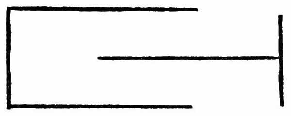
The first thing we learn when we study light is that it travels in straight lines. The chief source of light to the inhabitants of this earth is the sun. Now the sun is so far away that, for all practical purposes, the rays of light coming from it may be looked upon as being parallel to one another. That[41] we must always remember, when dealing with the sun, though, of course, it does not apply when we are dealing with lights near at hand, unless they are specially constructed to throw parallel beams or rays, whichever we elect to call them. To prove that light travels in straight lines is not difficult, and we may devise a number of experiments for the purpose. The doors and ventilators of many dark rooms, in which photographic operations are carried on, are constructed on the assumption that light cannot travel round corners. An arrangement as shown in the diagram will allow air, but no light, to pass. If light were capable of going round corners, some other arrangement would have to be devised for the ventilation of dark rooms.
Having learned so much about light, we come to the most important fact of all, as far as the action of light concerns microscopic work. When rays of light travel, from a substance like air into a substance like water, they are bent out of their straight course. Without any desire to introduce a number of unfamiliar words, we may venture to remark that, any substance through which light passes is called a medium. Some media are clearly more dense, more compact or solid—dense is the proper[42] word—than others. Water is more dense than air and glass than either. The bending of light rays is known as refraction. So now we may state our second law a little more concisely, thus:—When light passes from a medium into one more dense, or vice versa, it is refracted, and the more dense the medium into which or from which the light passes the greater the refraction.
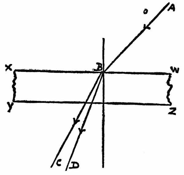
A diagram and an experiment should make matters clear. Suppose AB is a ray of light traveling in air and that it falls on a sheet of water, WXYZ, the ray will be bent along BC and its course from air to water may be represented by ABC. Suppose again, WXYZ represents, not water but glass; as glass is more dense than water the course of the ray AB is represented by ABD, it is refracted[43] or bent to a greater extent than the ray which passed from air into water.
For our experiment we need only plunge a stick into water and notice that, owing to this property of light, the stick appears bent, from the point where it comes into contact with the surface of the water.
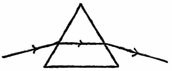
Some of us may be old enough to remember that once, on either corner of nearly every mantlepiece, there stood an ornament of doubtful utility from which there hung a dozen or more glass prisms. Now the only beauty about these otherwise hideous contraptions was to be seen when light played upon them. Then patches of violet, green, yellow and red were thrown upon neighbouring objects. White light, ordinary sunlight that is to say, is really composed of various colours—violet, indigo, blue, green, yellow, orange and red—which, when combined together, make light as we know it. When white light passes through a prism of glass, it is not only bent out of its course, but broken up into all these colours. A prism, as we all know, when examined at either end, is seen to be triangular in shape. Putting aside for a moment the question of the breaking up of light into its component parts, the path of[44] a ray of light through a prism is shown in the diagram. As the ray passes from air into glass it is bent, because glass is more dense than air; it is bent once more on leaving the prism because air is less dense than glass.
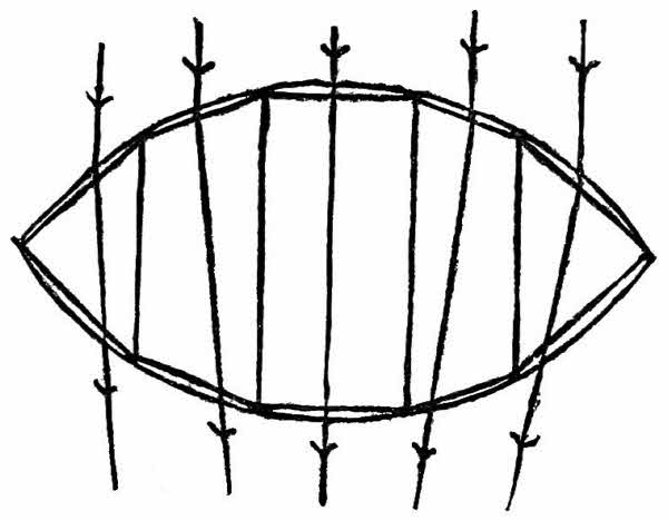
Now lenses are made of various shapes, and those with two outwardly curved surfaces are known as double convex lenses. A double convex lens is usually made with both its surfaces equally curved and in the finer optical work great care is taken to ensure that this is the case. For certain purposes, however, as we shall learn in a moment, one or other of the faces only may be much more curved than its companion and this may be carried to such an extreme that one face is flat, the lens is then known as plano-convex. Lenses may also have inwardly curved faces, if both are of this design they are called double concave; if one face is flat and the other inwardly curved they are known as[45] plano-concave. There are other combinations, for example, one face may be inwardly curved and the other outwardly curved, but the four kinds we have described are all that need trouble us.
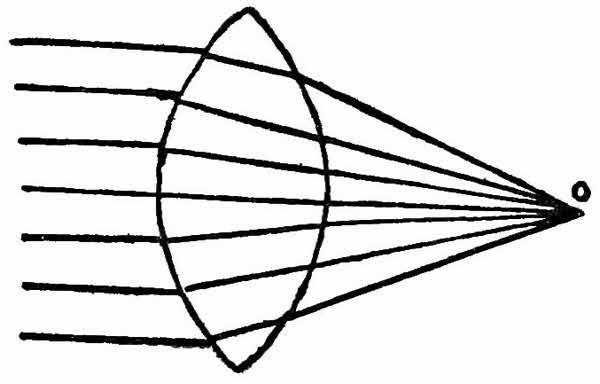
It does not require a great amount of imagination to recognise that the double convex lens, that is the lens with two outwardly curved faces is little more than a pair of prisms placed base to base, or more accurately, a number of prisms so arranged as shown in the diagram. Parallel rays of light falling upon such an arrangement of prisms would be bent from their course, as shown by the arrows, and this is just what happens with a double convex lens. Now rays of light from an object, passing through a lens of this shape may follow any one of three courses, according to the position of the object with regard to the lens. In one position and one only the rays after passing through the lens will be parallel to one another, as shown in the diagram.
The only position of the object for the above to take place is when it coincides with a point known as the principal focus of the lens, conversely the[46] parallel rays of light from the sun, after passing through a double convex lens, will come to a point at its principal focus.
Suppose now that the object be placed at a point beyond the principal focus of the lens, the light rays therefrom will, after passing through the lens, converge to a point thus:—
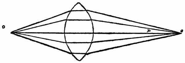
In the diagram O is the object and P the principal focus of the lens.
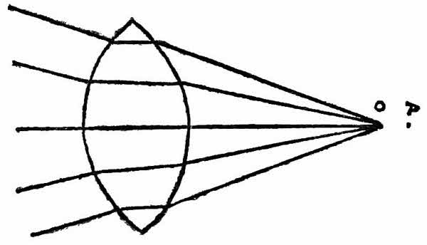
The third case occurs where the object is nearer to the lens than its principal focus, then the rays after passing through the lens, diverge and never meet.
We have already stated that when white light passes through a prism it is broken up into different[47] coloured rays varying from violet to red. The reason for this is that all the light rays composing white light are not bent equally as they pass from one medium to another. The violet rays are bent the most, the indigo next, blue next, down to red, which is least bent. Once more, considering the double convex lens as made up of a number of prisms, let us represent, by a diagram, the course of parallel rays of white light through it.
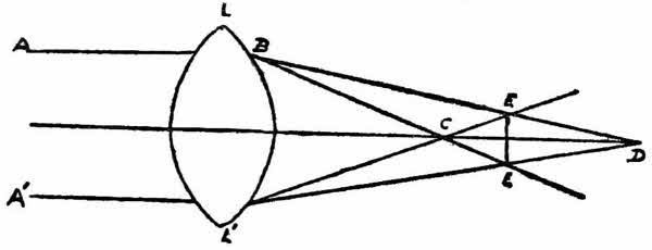
A A′ represent the parallel rays of white light falling on the lens, L L′. The blue rays are bent more than the red, so the principal focus of the former is at C and of the latter at D. The consequence of this difference in bending of the various coloured light rays would be most serious in microscopic work were not means devised to overcome it. Objects for instance at C in our diagram would appear blue, at D they would appear red, whilst at E E′ though no single colour would predominate they would be illumined with many coloured rays, though less strongly than at C or D.
This chromatic aberration, as it is called, depends amongst other things on the nature of the glass used in lens construction. It has been found, however,[48] that a combination of flint and of crown glass will overcome the difficulty. In a later chapter we shall explain the difference between these two kinds of glass. In practice, a plano-concave lens of flint glass is combined with a double convex lens of crown glass and, if the nature of the glass is satisfactory, as also the shapes of the lenses, there is full correction for chromatic aberration, and objects viewed through such a lens will not appear with coloured margins.
There is one further trouble likely to occur in such, or any lens. We write of the rays meeting at a point. In our diagrams we represent the rays by straight lines, really they are much more complicated than they appear in a diagram. It is quite easy to take a ruler and make our imaginary light rays meet at a point, as a matter of fact, where real lenses and real light rays are concerned, it is very difficult, if not impossible, to make the latter meet at a single point. One more diagram may make the matter clear.
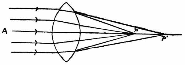
Parallel rays A pass through our lens and, as we know, they should all meet at a point P, the principal focus of the lens; the majority do so, but[49] some meet at other points, such as P′. In consequence of this it is difficult to obtain a clear image of an object at P, and the lens is said to suffer from spherical aberration. The perfect simple lens would be one fully corrected for chromatic and spherical aberration.
In our chapters, dealing with, the history of the Microscope, we attempted to trace the gradual development of the compound instrument from the simple lens; we stated that the latter, in a crude form, had been known and used from very early times and that the former developed side by side with the telescope. We have also said a few words in Chapter III. concerning light for the reason that the microscope can be better understood and used more efficiently when we are acquainted with the phenomena due to light. The simple lens, sold under the name of pocket magnifier, in its cheapest form consists of a double convex lens, that is to say, a lens with two outwardly curved surfaces. Better quality pocket magnifiers consist of two or more lenses, which may be either double convex; plano-convex, i.e., with one surface perfectly flat and the other outwardly curved, or they may be constructed of a combination of double convex and plano-concave lenses, such as were described on p. .
The object of both the simple and compound[51] microscope is to make objects appear larger than they do to the naked eye. When we buy our pocket lens we shall find that these little instruments are constructed to give different degrees of enlargement, some make objects appear five times larger than they do to the naked eye, some ten, some fifteen and some twenty times larger. Twenty times is about the limit of magnification for the ordinary pocket lens. If we are observant we shall notice something else—the greater the magnification the nearer we must hold the lens to our object. Within certain limits, this is not a very serious matter, but a point is reached where we must hold our lens so near to the object that we cannot see it, and that is why we cannot obtain very great enlargement with a pocket lens. Despite this fact, as we read in our opening chapter, some very wonderful discoveries have been made with these simple microscopes.
Now we wish to show how a compound microscope works and, having done so, to explain the uses of its various parts. We shall consider the lenses of the instrument to be double convex; we do this for the sake of simplicity. Even in the cheapest compound microscopes of to-day simple convex lenses are never used, for the reason we explained in our last chapter. To understand the course of the light rays passing through our microscope, however, we may look upon the lenses as being merely double convex.
Let us try a simple experiment first of all. For the purpose we require two double convex lenses,[52] one capable of magnifying more than the other, a sheet of paper and a candle. We must darken the room in which we make the experiment and, having lighted the candle, we may proceed to make a compound microscope, for that is really what we are about to do. Taking the lens which gives the greatest magnification, we look through it till we can see a clearly defined image of the lighted candle, then we fix the lens at that spot, so that, during the rest of our experiment, the candle and lens remain at the same distance from one another. Now we put the piece of paper as nearly as we can in the position of our eye, moving it nearer or further from the lens till we have a perfectly clear image of the candle thrown upon it. The first thing to strike us is that the image is upside down; it is known as a real, inverted image. Real because it can be thrown upon a screen and inverted—well because it is upside down. There are some images, as we shall learn in a moment, which can be seen but which cannot be thrown upon a screen: they are called virtual images.
Having fixed our sheet of paper in position, we take our second lens, focus it sharply upon the back of the sheet of paper, being careful to keep the centres of the two lenses as far as possible in a straight line with one another. Having obtained a sharp image we remove the paper and gradually advance our second lens towards the first. We soon reach a point where we have a very much larger image of the candle than the first lens gave us; we[53] must fix our second lens at this point for we now have a compound microscope, a very crude one certainly and without the trimmings which make the microscope so useful. Before we proceed to explain what has happened to the light rays we must take our paper screen once more and place it as near as possible to the spot where our eye was situated when we saw the second image. We shall find that, however much we may move our screen to or from the second lens we can never manage to obtain an image upon it for the reason that this second image is virtual, but unlike the first image it is not inverted.

One or two diagrams will help to explain our experiment and, instead of the lighted candle, we will suppose that our object is an arrow—it is easier to draw and serves just as well. The magnification of the object by our first lens may be represented by the diagram below, where AA is the lens, CD the object and D′C′ its image.
The arrow C′D′ shows the point at which we placed our screen, and as our diagram shows, the image is magnified and inverted.
Our second lens, we remember, was focussed on the back of the paper, placed at C′D′; for practical purposes we may ignore the thickness of the paper and say that it was focussed on the image C′D′. Had we left it at that, the further course of the[54] rays through the second lens would be represented by a replica of the diagram we have just given. But, in our experiment, we moved the second lens nearer and nearer to C′D′ till we obtained a clear much magnified erect image of C′D′, let us call this second image C″D″, and represent the course of the light rays by a diagram.
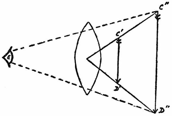
We may well ask, why did the lens AA, our first lens, form a real image whilst the second lens BB, which is precisely similar to AA, except that its magnifying power is not so great, form a virtual image? The formation of a real or a virtual image is nothing to do with magnification, so we repeat—why do two similar lenses form different kinds of images? Let us refresh our memories with the remarks concerning the principal focus of lenses in the last chapter, then we may try another experiment. The principle focus of a double convex lens, we remember, is the point to which parallel rays[55] of light converge, after passing through the lens. If now our object is further away from the lens than its principal focus, a state of affairs that existed in the case of our lens AA and the object CD, we obtain a magnified, real but inverted image; if, on the other hand, using the same lens if we wish, the object is nearer to the lens than its principal focus, we obtain a magnified virtual and erect image. The form of image then depends on the relative positions of lens and object and not on the magnifying powers of the former.
After this digression, we will see what happens when we combine the diagram showing the real, inverted image, formed by the lens AA with the virtual erect image, formed by the lens BB. In reality we will draw a diagram showing the path of the light rays through our compound microscope.
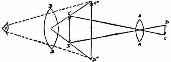
We have used the same lettering as in our previous diagrams and we see that, also as before, a real, inverted image C′D′ of the object CD is formed by the lens AA and a virtual, erect image C″D″ of the image C′D′ is formed by the lens BB, the object CD being further from the lens AA than its principal focus and the image C′D′ being nearer to the lens BB than its principal focus. One very important[56] point we must notice before we leave the diagram. We have mentioned several times that the image formed by BB is erect and so it is, but it is an erect image of an already inverted image, so that the final image of CD, as seen by the eye E is inverted. The fact that objects viewed through the microscope appear upside down is puzzling at first. To all intents our two double convex lenses represent a compound microscope; actually, they should be fixed at either end of a tube, blackened on the inside. The lens AA, nearest to the object, would then be known as the objective and the lens BB nearest to the observer’s eye would be known as the ocular or, more commonly the eyepiece. There are, of course, very many refinements, designed to make the instrument capable of performing the most accurate work, and needless to say these simple lenses would neither give very great magnification nor any clear images. Let us describe a more refined compound microscope than the one we constructed in our darkened room. The optical parts, that is to say the lenses, are the most important parts of every microscope, upon their qualities depend the degree of efficiency of the instrument; the metal portions, known collectively as the stand, contribute to the easier, smoother working of the microscope.
By the courtesy of Messrs. F. Davidson & Co.
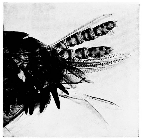
The Head of a Dog Flea
No wonder the flea is an annoying creature. As the plate shows, it is armed with knives, lances and saws, all designed to injure the skin of its victim.
The stand must claim our attention first. The base of the instrument, called the foot, is usually either three-legged or horse-shoe shaped; whatever its form it should be heavy, for only thus can the microscope be steady, and steadiness is essential in[57] all microscopic work. At the top of the foot there is a joint, in order that all the other parts of the stand may be inclined at any angle, from the vertical to the horizontal. Just above the joint is a bent arm of brass, to the forward end of which a brass tube is affixed. This tube is designed to hold the lenses, the objective at its lower end, the eyepiece at its upper end. The tube is always blackened inside; were this not the case, light passing through the objective would be reflected in all directions from the sides of the tube and a clear image of the object could never be obtained. The tubes of microscopes vary in length according to their country of origin; English and American tubes are ten inches long, those of continental make vary from a little more than six inches to rather more than seven inches in length.
Affixed to the lower end of the bent arm of brass, mentioned above, is a flat metal plate, known as the stage; at its centre, there is a circular hole through which rays of light pass to illuminate objects placed upon it. Below the stage, at the edge nearest to the foot, there is a metal peg, over which fits a tube to which a mirror is attached by a moveable joint. The mirror reflects light rays through the opening in the stage. The tube, holding it, can be slipped up and down the peg under the stage, thereby bringing it nearer to or further from the object and so altering the intensity of the reflected light, as we shall explain in a moment. Owing to its moveable joint, it is possible to swing the mirror[58] to the right or left, so that the reflected light rays do not pass directly through the object on the stage, but strike it on one side or the other, thereby giving what is known as oblique illumination.
The cheapest forms of compound microscopes have all the parts we have mentioned, and focussing is carried out by sliding the tube, with its objective and eyepiece, up and down within its holder, in order to bring the objective further from or nearer to the object.
In more expensive instruments there are further refinements, in fact, on some of the very costly present-day instruments, there are so many appendages and appurtenances that it is doubtful whether some of them are not more of a hindrance than a help, at any rate they increase the possibility of trouble by their liability to get out of order. Such microscopes are only of use to very expert workers; there are, however, a good many additional features to be found on quite moderate-priced instruments, features which are a great help to the microscopist.
It is obvious that we cannot attain any degree of accuracy in focussing, especially with high magnifications, when we must perforce raise or lower the tube by hand. To obviate this difficulty, most microscopes are provided with mechanism known as a coarse adjustment; it consists of milled screws at either end of a metal rod; in the centre of the rod there is a little cog-wheel which engages with a row of notches on the tube. By turning the milled screws slightly in either direction, we can impart[59] a considerable upward or downward movement to the tube carrying the objective and focussing at once becomes a more simple matter. The coarse adjustment is only useful for examining objects with a low magnification; if we use it when objects are being highly magnified we run the risk of screwing our objective down upon our object, to the certain destruction of the latter and the probable injury of the former. To obviate such a catastrophe, most of the better class microscopes are also provided with a fine adjustment. By means of this adjustment, which externally takes the form of a single milled screw, a considerable turn of the screw in either direction only imparts a very slight upward or downward movement to the microscope tube. In the best instruments, movements of as little as one hundredth part of a millimetre may be imparted to the tube by the fine adjustment and, seeing that there are about twenty-five and a half millimetres to the inch, it is obvious that a good fine adjustment is very delicate and, being so, must be treated with care. The fine adjustment is used to supplement coarse adjustment in the final focussing, when using high magnifications.
A few words may be devoted to the mirror, for on its intelligent use much depends. Usually we shall find that it is plano-concave, that is to say, flat on one side and hollowed out on the other. The use of the mirror, as we have mentioned already, is to reflect rays of light through the opening of the stage on to the object we desire to examine. Both[60] mirrors will reflect parallel rays of light to a point, just as a double convex lens will so direct them from their course that they meet at a point. The concave mirror gives the more powerful illumination, because it reflects more light rays than a flat mirror of the same diameter.
We have mentioned that, to obtain full advantage from the mirror it should be capable of movement to and from the stage. When we desire strong illumination we arrange the mirror so that its reflected rays meet at a point coinciding with our object. Should less intense illumination be required, we slide the mirror nearer to the stage, and of course nearer to our object, so that the reflected rays meet at a point above the object.
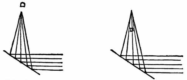
The two diagrams, given below, show the path of the rays of light, where O is the object, and a trial with our microscope will soon show which position gives the more powerful illumination.
For high-power work, such as bacteriology or even the examination of sections of plants, etc., even the best concave mirror will not give a sufficiently powerful illumination; accordingly an instrument,[61] known as the condenser, is fixed below the stage, between the mirror and the object. The condenser, as its name implies, condenses the rays of light reflected to it by the mirror. It consists of a series of lenses so arranged that they will throw a very powerful cone of light. Provision is made for focussing the rays from the condenser on to the object.
Sometimes, for special forms of illumination, it is necessary to cut off some of the rays of light passing through the condenser. It may be that we desire to dispense with the outer rays of the cone of light or, when delicate details are being studied, we may wish to impede the central rays. In either case diaphragms, popularly called “stops” are used. Our diagrams show A the outer rays of a cone of light cut off and B the central rays similarly treated.
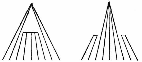
In old pattern microscopes and in many instruments not provided with condensers, the diaphragm used for the purpose of cutting off the outer rays of the cone of light, consists of a blackened circular metal plate, perforated with a number of different sized circular holes. This plate is fixed below the[62] stage in such a manner that, as it is revolved, holes of various diameters are brought one by one within the cone of light. It need hardy be remarked that the smaller the hole in the diaphragm the more light is cut off and the less reaches the object. In more modern instruments and in practically all which are fitted with a condenser, an Iris diaphragm is fitted. A diaphragm of this nature consists of a number of thin, blackened, metal leaves, fastened to a metal ring in such a manner that, when the ring is revolved, the leaves close together, making the opening in the centre smaller and smaller. The Iris diaphragm has many advantages over the old perforated metal plate. At will, we can have any opening from full to the merest pin-point or we can cut off the light rays altogether, should we wish to do so; we are not confined to a definite number of stops. As we cut off these outer rays of light we shall find that, up to a certain point, though the illumination becomes less and less the object becomes more and more clear, or, to use the correct expression, its definition is improved.
When it is necessary to cut off some of the central rays of the light cone, either a circle of glass with an opaque centre is dropped into a metal holder below the stage, or a circular metal plate, held in the centre of a metal ring by three arms, is used in the same manner.
The effect of cutting off the central rays of the light cone is, of course, to reduce the illumination and to show up delicate detail to advantage. No[63] direct rays of light reach the objective, such as do pass into the microscope are all diffused from the edges of the object.
We have already mentioned that the optical parts of the compound microscope are of greater importance than what may be termed the mechanical portions and the objectives are more important than the eyepieces. Better results can always be obtained with a good, high-power objective and a low-power eyepiece, than with an inferior objective and a good quality eyepiece. The merits of the eyepiece, however great, will not be adequate compensation for the failings of the objective. Modern objectives are composed of several lenses and of a combination of flint and crown glass, as we explained in our last chapter. They are so designed that they can be screwed into the lower part of the microscope tube. The focal length of each objective is, or should be, marked upon it; as a general rule, however, it may be taken that the smaller the lower lens, the shorter its focal length and therefore the greater its magnifying power.
The form of eyepiece most usually met with is known as Huyghen’s. It consists of two plano-convex lenses, with their flat or plane surfaces directed away from the objective. The smaller of the two lenses is situated nearer to the eye of the observer and is known as the eyeglass; its function is to magnify the image formed by the objective. The larger, lower lens is known as the field or collecting glass; it renders the image clearer though, in so[64] doing, it reduces the magnification of the eyeglass. In instruments provided with more than one eyepiece we shall wish to know which gives the greater magnification; this is or should be marked upon the metal rim surrounding the eyeglass but, in general, it may be stated that the shorter the eyepiece the greater its magnification. We repeat again, increase your magnification always, when possible, by using higher power objectives rather than eyepieces with greater magnifying powers. Sometimes it is necessary to use a greater magnification than our most powerful objective will give us; then we must fit our most powerful eyepiece and draw out the upper part of the microscope tube—in the best instruments they are made to pull out, after the manner of the telescope. The effect of so doing will be to increase the magnification considerably but, at the same time, the definition or clearness is seriously impaired.
For the examination of practically all our microscopic objects we require a number of slides, little glass slips of good, thin, clear glass. They may be used over and over again unless we make permanent preparations, but we are hardly likely to do so in our early days. The slides are held in place on the microscope stage, either by a pair of clips attached thereto or by resting against a bar running across the stage. We may here remark that it is essential always to keep one’s microscope slides absolutely clean. Dirty slides denote the careless worker; moreover, dirt when magnified is misleading. Objects which are being examined in water or any[65] other liquid should be covered with a cover-slip, an exceedingly thin circle or square of glass. The cover-slip is as much a protection for the objective as for the object and its cleanliness also, is all important.
We have not mentioned any refinements such as the mechanical stage, by means of which slides on the stage may be rotated, moved to the front and to the back of the stage or from side to side. We have omitted these because they are not essential even for the very best work; they lend additional comfort to the use of the microscope but, again, they are not essential. The microscopist who requires such luxuries may learn about them in the larger text-books on the microscope.
The enthusiastic microscopist will probably never lack material for his instrument, whatever branch of microscopical work he may decide to make his own. To the student of Pond Life, either animal or vegetable, there is granted a never-ending store of beautiful and interesting objects. Because one pond has been thoroughly searched and all that it can offer has been carefully examined, we must not conclude that no other pond will be worth our attention. Though indeed many little animals occur over and over again in practically every pond, there are other equally interesting animals which only occur in certain localities and for these we must keep a sharp look-out.
The apparatus needed for the collection of the denizens of ponds and streams, need consist of no more than a net with a very fine mesh and a jar[67] in which to bring our captures home; for, of course, animals which dwell in water need not be dried on the journey home. Various useful accessories for the student of pond life are sold at very reasonable rates by most scientific instrument makers.
We shall find many representatives of the animal world in our pond and exceedingly interesting most of them will prove. From the mud we may obtain the “protean animalcule,” known to scientists as Amœba Proteus, the most lowly of all animals. Though this creature is plentiful and just visible to the naked eye, he is not easy to separate from his surroundings. He is almost colourless and therefore paler than the mud. Having secured him on the end of a glass rod, let us examine him in a drop of water on a slide. At first he will remain motionless, as a protest against being disturbed; we shall not have to wait long, however, for soon one part of his body will be seen to protrude and then grow larger and larger till it forms a false foot; other parts may follow suit, till he is more elongate than oval, and he moves in the direction of his false foot with a curious gliding motion. His pace is not great and has been calculated at a twenty-fifth of an inch in an hour. Really the “protean animalcule” is little more than an animated drop of jelly, a fact we can substantiate by watching him feed. His food consists of minute water plants such as diatoms, and when one of these plants comes within his line of march he simply surrounds it with his false feet and, as it were, flows around it. When he has[68] digested all he can he flows away from the undigested portions; he has no mouth or any of the organs usually associated with animal anatomy.
While hunting for our Amœba, it is highly probable that a very active little slipper-shaped organism may have forced himself upon our attention. From his shape he has earned the popular name of the slipper animalcule. He is rather more highly organised than the Amœba for he possesses a mouth, as we shall see when we are able to examine him. So rapidly does he swim, however, that something must be done to curb his activity; he may either be killed with a drop of weak acid or we may put a little tuft of cotton wool on our drop of water and a coverslip lightly over that. The threads of cotton wool will form a network, in the meshes of which the active little animalcule will be confined. Careful observation will show that he is, like a slipper, more pointed at one end than at the other; that there is a funnel-shaped orifice, his mouth, at one side of his body; and that he is covered with little threads which lash the water with rhythmic movement and propel him with considerable rapidity. These little threads also send currents of water to his mouth, and in the water is his food.
Having examined these two free swimming denizens of the pond, we may advantageously turn our attention to some of the weeds growing therein. Careful examination with our pocket lens will almost certainly reveal a number of minute living creatures attached to the submerged stems and leaves. It[69] is impossible to describe all the interesting creatures we might find: we must content ourselves with two, because they are common inhabitants of many ponds. One interesting little creature we are almost certain to find, the Hydra. He may be green or he may be brown but in structure he will remind us of a small sea anemone. When we put him under the microscope he has the appearance of a mass of jelly attached to the water plant on which we found him. He will soon open himself out, however, and we shall see that his free end is provided with a number of tentacles; these he waves about in the water to draw small swimming creatures to his mouth which is situated in the centre of the group of tentacles. Any luckless creature, coming within reach of the Hydra, is at once stung with one of the barb-shaped stings which stud his tentacles and then passed to his mouth. The Hydra is wonderfully tenacious of life; it is said that he has been turned inside out, like a glove finger, without suffering any inconvenience. Probably the specimen we are examining will have a swelling on the side of its body, which might be mistaken for the result of some injury; it is nothing of the kind; it is merely a bud which will grow into a young Hydra and, when old enough, become detached from its parent and float away to another plant. Under a fairly high magnification, we shall almost certainly see something gliding rapidly over the body of the Hydra; its movements are too quick to allow of careful examination; the creature which is almost as elusive[70] as the slipper animalcule is a parasite of the Hydra.
Not quite so common as our last object but still common enough to be mentioned here is the beautiful “bell animalcule.” Like the Hydra, this creature, except in its very young stages, remains affixed to a water plant. In shape the “bell animalcule” resembles a wineglass on a long delicate stem; round the part corresponding to the rim of the glass, there is a fringe of the hair-like, water-lashing structures with which so many of these lowly creatures are provided, and these structures also surround the entrance to the funnel-shaped mouth. When undisturbed, the bell animalcule has its slender stalk fully extended and its little threads lash the water vigorously, causing currents, containing food material, to travel towards its mouth. A sharp tap on the microscope slide will cause the creature to contract, the threads cease their lashing and the stalk contracts spirally, so that the body of the animalcule is drawn close to the object to which it is attached. By degrees the spiral uncoils and the little threads resume their lashing.
Sometimes, as we examine our bell animalcule, we may be fortunate enough to see it splitting into two parts to form two separate individuals. This curious process should be watched carefully. The upper part of the bell splits first and, by degrees the whole bell divides into two equal parts so that we have a pair of bells on a single stalk. The next stage consists of the formation of a ring of whip-like structures round the base of one of the bells;[71] both bells, by the way, have the circlet of whips round their upper edges. Soon after these additional little whips are formed, the owner of them breaks away from the stalk and swims about in the water for a time, finally coming to rest on a suitable water weed. Then the lower ring of whips has served its purpose and in its place a long stalk grows; from this time forward the new bell animalcule will never move from the position it has chosen. This form of increase, this simple splitting takes place over and over again but by degrees the little animal appears to become exhausted and the process slows down or stops.
The partially exhausted Vorticella may gain increased vitality by fusion with another individual and this process also we may have the luck to see though it is less frequent than the simple splitting. Sometimes a bell may be observed to divide, not into halves, but into two unequal parts. The smaller of these parts may divide again into from two to eight parts, each one of which, having developed a fringe of little whips, swims off on its own account. These little barrel-shaped swimming forms, instead of settling down and forming stalked bells, seek an exhausted creature, fuse with it near the base of its bell and finally become absorbed by it. The result of this fusion is that the bell animalcule takes on a new lease of life and once more begins to divide actively.
In our search for specimens for our microscope we may come across a very common pond dweller,[72] closely related to the bell animalcule, known by the name of Carchesium Spectabile. We cannot fail to recognise its family likeness to the form we have already studied, for it consists of a large number of stalked bells growing on a single parent stem. It is really a little colony of bells. When one of the young individuals of Carchesium settles down in the spot it has selected for its dwelling-place, it grows a stalk just as Vorticella did, and it divides later into two individuals. Now in Vorticella only the bell divides, in Carchesium part of the stalk divides also and, instead of swimming away to find a new home it remains attached to the parent stalk. When this has happened several times a goodly colony is formed.
There are few fresh-water animals more commonplace and apparently uninteresting when observed casually than the pond sponge and the river sponge. Yet, if we take either of them home and examine them with the aid of our microscope, we shall be delighted with our specimens. In reality they are of absorbing interest and, at certain times of the year we may easily obtain young sponges, and capital objects they make for the microscope.
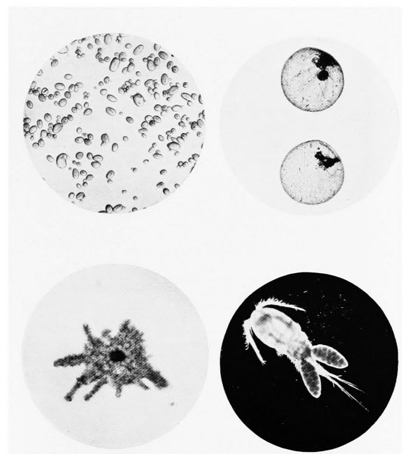
Photos by Flatters & Garnett
1. Starch Grains of Potato
Starch is largely used as an adulterant of various foods. The Potato starch grain resembles a miniature oyster shell.
2. Phosphorescence Animalculae
These minute animals, occurring in millions, render sea water beautifully phosphorescent.
3. Proteus Animalcule
The lowliest of all animals. It is a jelly-like animal which changes its shape from hour to hour.
4. Cyclops
A common one-eyed water animal. The female, illustrated, carries her eggs in a pair of relatively large bladder-like sacs.
Before we describe our two specimens let us try to explain what manner of creature a sponge really is. If we examine any bath sponge, we notice that it is perforated with many small holes and some larger ones. Some sponges show this better than others. The small holes are pores, the large ones mouths, but, as we shall see in a moment we must[73] not run away with the idea that they in any way resemble our familiar idea of a mouth. These large holes are called oscula by scientists, but we wish to avoid scientific words as much as possible. The simplest sponge of all, consists of a little bag, which remains affixed by its base to a seaweed. All over its sides there are many pores and at its tip there is a single osculum; it is known as the Purse Sponge and is common round our coasts. The inside of the purse sponge is lined with cells, each one of which is tipped with a little whip which waves about unceasingly. The waving of the whip causes water to flow through all the pores into the hollow bag and out again by way of the osculum. Although most sponges, including our fresh-water forms, are much more complicated than the purse sponge, the same thing happens in them all, water is drawn in by way of the pores and forced out by way of the oscula.
The best place to seek for the fresh water sponges is on the under sides of floating wood, broken tree branches and the like. Their appearance depends upon whether they have been growing in a well-lighted spot or in darkness; they contain chlorophyl as do the higher plants, and sponges grown in the light are green, those which the light has not reached are buff-coloured or corn-yellow. The pond sponge is brighter green than its river frequenting relative and is a coarser creature altogether. It often forms little finger-like outgrowths, whereas the river sponge is more leaflike.
If we examine one of these sponges in a watch glass of water to which we have added a very little carmine powder, we can easily see the water currents entering the pores and coming from the oscula. Towards autumn, if we open a river sponge we shall see little yellow bodies about the size of a pin’s head; they are the buds from which the young sponges arise. One of these must be removed very carefully and placed in a watch glass of water; if our specimen be placed in the sun, we shall not have many days to wait before we find that it has given rise to an active transparent little creature, to a young sponge in fact. By repeating our experiment with carmine, by the aid of the microscope, we can see the water currents passing through its body.
Wherever we collect our pond water we are certain to find some of the wheel animalculæ or rotifers. There are such a number of different kinds that we might describe several, and yet not mention the one that any of our readers had happened upon. They may be recognised, because they are always so transparent that all their internal organs may be plainly seen and they always have two or more discs or lobes, at their forward end, fringed with fine whip-like threads. These little threads are constantly in motion, so that they appear like little revolving wheels hence the name, wheel animalcule. Nearly all these creatures have a sucker or false foot at their tail end by means of which they attach themselves to some support or they may swim freely in the water. The study of rotifers has been[75] the life-work of some scientists and they will afford any microscopist, who is interested in them with abundant occupation.
Amongst the water weeds we shall probably find another small but striking creature, known as the sun animalcule, Actinophrys Sol. Why exactly it is called the sun animalcule we cannot say, probably it has earned its title from the fact that it resembles the conventional idea of the sun, with light rays radiating all round its circumference. The creature is whitish-grey in colour, spherical in shape and just visible to the naked eye. All over the surface of its body there are a number of apparently empty spaces, known as vacuoles—they probably account for its peculiar colour. Everywhere, it is studded with long, slender-pointed rod-like outgrowths. But rarely, the sun animalcule exhibits any movement and for long periods the only signs that it is living occur at feeding time. Its food consists of water animals and plants, of varying size. When a small animal comes into contact with one of the pointed rods, which radiate from the animalcule, it appears to be held there in some unaccountable manner and, after a pause, it begins a fateful journey by sliding down the rod to the spherical body of its captor. Then it is passed into one of the vacuoles and digestion very soon takes place. That it does not do so immediately is shown by the fact that the wheels of a wheel animalcule, which has been passed to the vacuole of our subject, continue their movements for a considerable period. When larger[76] animal food is partaken of, a different method is pursued by the sun animalcule. A water-flea, for instance, coming into contact with one of the rods will struggle violently in its efforts to escape. Then the sun animalcule shows real signs of life, for some of the other rods bend over and hold the captive so that it, eventually, is passed to a vacuole.
In our chapter on agriculture we mention a peculiar flat worm known as the liver fluke. This unpleasant creature has one or two relatives who make their home in ponds and, though they are not more beautiful than the liver fluke to look upon, they are quite harmless. One of these flat worms is about 3/4-inch long and not unlike an indian club in shape. It moves about, with some speed either by the aid of a sucker on its head or by a curious gliding movement. The other pond-frequenting flatworm is more like the liver fluke, its leaflike oval body, about 1/2-inch long, is pointed fore and aft. It is a common sight to see it gliding here and there in search of still smaller animals off which it may make a meal.
The sea mat and bird’s head, two common animal colonies of the sea-shore, possess pond-dwelling relatives of the greatest interest to the microscopist. Like the seaside forms they dwell in colonies. One of the commonest is known as Lophopus Crystallinus and it may be found attached to duckweed, the curious little plant whose tiny leaves float upon the surface of the water. Lophopus, when at rest, resembles a little piece of jelly. If we are patient[77] and watch it under our microscope we shall see it expand, sending forth a number of stalks, each one tipped with a horse-shoe shaped feathery tentacle. Each of these tentacles belongs to a separate animal which with its fellows forms a colony. Not so interesting are the branched, threadlike colonies of Plumatella Repens which may be sought upon the leaves of water plants. Our ponds can furnish no more extraordinary object for our microscope than a colony of Cristatella Mucedo; it is curious in appearance and still more curious from the fact that though a colony of animals it acts like a single individual in crawling over the weeds and stones in shallow, sun-kissed water. The Cristatella colony is jelly-like and greenish in colour, in length it may grow to a couple of inches. Its under surface is flat, whilst from its upper, convex surface the little animals forming the colony wave their brush-like tentacles in the water.
In searching the various pond weeds for specimens we are sure to meet with various jelly-like masses; these must always be examined carefully. They may be the egg-masses of interesting water creatures; various water-snails, for instance, protect their eggs with a jelly-like covering.
If we meet with any large fresh-water mussels, sometimes called swan mussels, we shall probably find the fleshy parts of the molluscs swarming with minute creatures, which we may conclude are parasites. Mussels, like nearly all living creatures, have their parasites it is true, but what we have discovered[78] will almost certainly prove to be young mussels. We must examine them under the microscope to make sure. Then we shall see, if young mussels they be, very minute and very thin-shelled little creatures; the edges of their shells are armed with teeth and are quite unlike the highly polished and smooth shells of the parent mussel. We shall also see a long thread issuing from the animal within the shell. If we are examining the young mussel in water we shall notice that it is constantly snapping its two shells together.
Had we left these young mussels undisturbed, a very curious life they would have led. They would have remained attached to their parent for some little time probably, or some of them might have fallen to the muddy bed of the pond. Their behaviour in either case would be the same. The long threads that we have already examined would have floated in the water and, directly they were touched by a passing fish, the little shells would begin to snap violently. The lucky ones would not snap in vain for they would close upon the fin or tail of the fish and then their snapping would cease. Like the bulldog, these little mussels may take a long time in getting hold but when once they have managed to fasten their teeth into anything it is well-nigh impossible to make them let go. Usually they never let go, but are carried away by the fish. In most cases, the irritation they set up in the flesh of their new-found foster parent causes a swelling to occur with the result that the little mussels are[79] engulfed. Within the flesh of the fish they go through a series of changes; they lose their teeth and their tell-tale thread, they become in fact miniatures of the adult mussels, then they manage to escape from the fish, settle down in the mud and fend for themselves.
The crabs and lobsters which we know so well have fresh water relatives in nearly every pond. Many of the creatures we have examined have needed careful search to discover their whereabouts; not so the fresh water crustacea, as they are called. Their activity, their curious movements in the water compel attention.
The fresh water shrimp is a curious little creature, sometimes he paddles his three pairs of hind legs and sometimes he jerks his body in a ludicrous manner, in either case he manages to propel himself rapidly through the water. He is about half an inch long, brown in colour and with a curved body not unlike a shrimp. If we examine him under the microscope we notice that his front legs are bent forwards, whilst his hind legs are bent backwards. The male water flea is much larger than the female, a fact which probably accounts for the fact that these little animals often carry their wives about with them by seizing them with their fore legs.
The water louse we may also encounter, he is not nearly so interesting as the fresh water shrimp. He is closely related to the wood louse which we all know, and has a similar flattened body.
Very much smaller though even more interesting[80] is the common water flea. By day these animals retire to the mud at the bottom of the pond but, morning and evening, they swim actively with a curious jerky motion. We must examine our specimen carefully for he is of more than ordinary interest. We cannot fail to observe how transparent he is, so much so that all his internal organs can be plainly seen, but let us deal with his exterior first of all. His large eyes are plainly visible, but his most conspicuous feature is the pair of large branched feelers, by means of which he swims. If we examine several specimens, one or more is certain to be a female, then we may probably observe an egg in process of formation in the brood pouch, a large, elongated cavity, just below the back of the animal. Immediately above the brood pouch, the heart is situated and, if we can induce its owner to keep still for a moment or two, the heart-beats may be plainly seen.
Not unlike the water flea as it swims about in the water of our collecting jar is the curious, transparent little creature known as Cypris. Although so transparent its body is contained in a pair of shells, very similar to those of the mussel; a fact which formerly led to its being classed with the shell-fish. We may well examine this little fellow under the microscope for much of his structure may be made out through his shell. His very conspicuous eye is sure to attract our attention; he possesses but a single eye and seems to make up for the lack of a second by having a very large one. Two pairs[81] of feelers project beyond the shell in front. Of his two pairs of legs the foremost, or at least their tips, hang down below the shell, but the last pair, as we can see through the shell, are turned upwards. At the hinder end of the body, there are two long bristles, they may best be seen when Cypris is swimming. Of what use exactly these bristles may be to their owner is not definitely known, but it is thought that they are of service in keeping his shell clean.
Another active little animal, quite as common as the water flea, is sure to attract our attention. It is no larger than the water flea but much more elongated; some specimens are bigger than others, and the bigger ones are the females. Its name is Cyclops and, though so common, it has no popular name.
Cyclops is so named on account of the fact that it possesses but a single eye; it is, however, rather an interesting creature in other respects, so we will study it more closely. Looking down upon the creature, we see that the front part of its body is composed of an undivided shield, behind which there are four plates and behind these again there are in the male five and in the female four segments or rings, at the extreme tip there is a forked tail, each fork being furnished with a number of bristles. On the head are two pairs of organs, one pair long the other pair short and, if we observe Cyclops in the act of swimming, we shall see that the long pair of organs play the chief part. In the centre of the[82] front of the head there is a black or red patch—the eye. Very frequently we may meet with a specimen carrying a relatively large bladder-like body on each side of its abdomen. These bladders which are each about one-third as long as the creature which carries them, are egg-sacs.
If we are able to secure one or two specimens with egg-sacs attached, we can study the young Cyclops without much difficulty. Take a small glass tube—a test-tube as used by chemists will serve admirably—partly fill it with pond water and add a little water weed, then introduce the egg-bearing females and place in the light. We must watch the tube from day to day, and before long it will be evident that the young ones have arrived in the world, for we shall have no difficulty in seeing dozens of little white specks swimming about in the water and settling on the sides of the tube. We must remove one carefully on the end of a glass rod or on a paint brush and examine it in a drop of water under the microscope. This young creature is totally unlike its parent, it is oval and possesses three pairs of stiff bristles, of which the first pair are simple and the other two pairs are branched. Although the bristles are used solely for swimming at this stage, it may be of interest to mention that in the adult Cyclops they become transformed into jaws and the two pairs of organs we have already examined. At the front end of the oval body we can closely distinguish the single eye, which persists throughout life.
In this chapter we shall confine ourselves to the true water-dwelling plants, as distinct from those, such as the water lilies, which though never found growing on dry land, appear undecided whether they will be water plants or land plants. Looking at the matter from a more scientific point of view, all our pond plants will be much lower in the scale of development than the water lilies and other flowering plants.
Pond life is rich in subjects for the microscopist. Any stagnant pool may contain organisms which will delight the naturalist who has always depended upon his unaided vision. Curiously enough, amongst the most wonderful of all these pond-dwelling plants are the Diatoms, which consist of but a single cell. They are so numerous, they exist in so many different forms and in so many different situations that were we able to describe them all, we should require the whole of a large volume, much larger than this. In colour, Diatoms are usually brown or brownish, although they contain chlorophyll, the green colouring[84] matter of higher plants. In shape they may be rod-shaped, crescent-shaped, circular, wedge-shaped, oblong or oval. Some float about freely in the water, some are attached to supports by means of stalks. Some lead a solitary life and others dwell together in colonies. One feature they have in common, a curious flinty cell wall, and this is their most interesting point to the microscopist. This natural armour is in two parts which fit one within the other like the two halves of a Japanese basket. All manner of beautiful sculpturing marks these beautiful frustules as they are called; in some cases they are perforated and the living matter from within passes through the pores and forms a jelly-like covering for the little plant.
We must make a point of collecting all the Diatoms we can find, for they are always interesting; moreover, they are easily preserved and made into permanent slides, for the little plants may be boiled in acid to destroy their living parts and the frustules will survive the boiling undamaged.
One might wonder how such humble plants, surrounded as they are with flinty walls, could increase. They frequently do so in a simple manner. The living matter of which the plant is composed pushes the frustules apart and divides across the middle. The result of this event is the formation of two plants, each with a single frustule. In a very short time each plant grows a new frustule, but it is always much smaller than the one with which it started.
The movements of some of the free swimming, that is to say non-attached Diatoms, are worthy of study. The scientific name of one kind, translated into everyday language, means little boats, and indeed they are well named for their beautiful aquatic manœuvres rival those of any ship.
Somewhat similar in habit to the brown Diatoms are the green Desmids, but, whereas, the former also occur in the sea, the latter are all confined to fresh water. Sometimes Desmids are so numerous that they make the pond water as green as green-pea soup. It would be as impossible to describe all these plants as was the case with the Diatoms, but generally they may be recognised by the fact that they are composed of two similar halves, separated by more or less of a waist. Although some of the Desmids exhibit a certain amount of movement they are not active like the Diatoms.
Late spring and autumn are the best seasons to hunt the ponds for our next object, which rejoices in the name Chlamydomonas Angulosa, a good example of the extraordinary fact that some of the smallest animals and plants have the longest names. This little plant is interesting in itself and doubly so, because it was for long thought to be an animal; it is wonderfully animal-like in its movements. Chlamydomonas is very minute, so we must use our highest magnification when we examine it. It is an oval, one-celled plant enclosed in a clear membrane. The green colouring matter is arranged in the form of a cup, within the hollow of which is a[86] mass of granular living substance. At the forward end of the plant, there is a clear space and near the tip a brownish dot, known as the eye spot; which, though incapable of seeing as we understand it, is sensitive to light, as shown by the fact that the little plant will swim towards moderately intense light and away from strong light. If we stain the plant with iodine we can plainly see a pair of little whips arising from the clear portion at the forward end; it is by the lashing of these that the plant is enabled to swim.
In the mud of our pond we may find a little colony of plants which might forgivably be mistaken for a collection of the individuals we have just studied. Each member of the colony is very much smaller than Chlamydomonas to be sure, but each one has the outer membrane, the brownish eye spot and the pair of little whips. On the other hand the chlorophyll fills the whole of each cell and is not arranged in the form of a cup. Sixteen cells form a colony, and the whole mass is a flat plate; the little whips move in unison and the whole colony revolves after the manner of a wheel.
Another little colony we may encounter also, consists likewise of sixteen cells very like the ones we have described, but somewhat wedge-shaped instead of oval. A jelly-like mantle encloses the colony and, in outline, it is spherical, so that when the little whips, which project through the mantle, lash the water the whole colony revolves.
The most remarkable of these colonies of cells is[87] known as Volvox Globator, it may be recognised by its perfectly spherical shape and its characteristic movements in water. Volvox is about 1/25 inch in diameter, and although to the uninitiated it appears to be a single minute plant, in reality it is a colony of upwards of twenty thousand cells. The colony may be considered as being made up of thousands upon thousands of cells, very similar to those of Chlamydomonas, and each one arranged with its pair of little whips directed outwards.
Within the Volvox sphere we may observe a number, usually one to eight, of smaller spheres. These are so-called daughter colonies which have arisen from the continued division of special cells. They develop, fairly rapidly, into young Volvox colonies, then they burst through the cells of the parent colony, swim out into the water and quickly grow to the size of the Volvox from which they were formed.
There is another kind of Volvox of a yellowish colour and much smaller than Globator, the big one is the one we must procure; it is much more easily studied.
Certain of the plants we shall find in our pond are so animal-like in their movements, that the microscopist who sees them for the first time may wonder whether we are not mistaken in calling them plants. We have already described the common Chlamydomonas, with its curious jerky method of propelling itself through the water. There is, however, an equally common one-celled, pond-frequenting[88] plant which has puzzled naturalists even more, for it certainly possesses many very animal-like characteristics. Its name is Euglena Viridis and we require our highest magnification to examine it for it does not exceed one-two hundred and fiftieth part of an inch in length. Usually, Euglena is cigar-shaped but, as it possesses the very unplant-like characteristic of not having a firm cell wall it can change its shape to a considerable extent and it often assumes curious forms. At the forward end of this minute plant there is a single whip-like thread, by means of which it swims; a little below the base of the whip, there is a red eye spot. Elsewhere we have described how the protean animalcule feeds by flowing round its food-material, Euglena feeds in a similar manner, but it also feeds after the manner of a plant. When this active little plant is about to increase it either divides into two lengthways or becomes surrounded with a firm wall within which it breaks up into a number of young forms which are released later by the bursting of the wall.
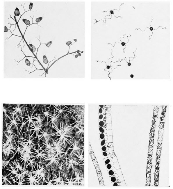
Photos by Flatters & Garnett
1. Bladderwort
A British water plant which entraps small animals in its bladders and digests its captives.
2. Spores of Horse Tail
These spores with their thread-like outgrowths vary in appearance according to the moisture in the air.
3. Hairs on a Potato Leaf
The star-shaped hairs on a potato leaf make beautiful objects for the microscope. Leaves of many plants are clothed with curious hairs.
4. Spirogyra
A green thread-like water weed. Two threads are shown fusing together; from each part of these fused threads a new plant will arise.
Small wonder that scientists were in doubt concerning the true nature of Euglena, its non-walled cell and peculiar mode of feeding are indeed puzzling. There are many similar cases amongst these lowly organisms, in fact there are some creatures which may best be described as partly plant and partly animal. Those of our readers who are interested in such problems may read an excellent account given much more fully than we could[89] give it here, in Professor F. W. Keeble’s Plant Animals, one of the excellent Cambridge Manuals of Science. The fact is that the demarcation between plants and animals, low in the scale of development, is not nearly so pronounced as it is amongst the higher forms of life. Amongst the plants of our pond we shall find that, like the land plants, most of them are green, and many of them are thread-like, so that the task of distinguishing one from another may appear difficult. Examined with the naked eye, many of them appear remarkably similar to one another; under the microscope the differences are obvious.
One of the most remarkable of the commoner pond plants is known as Oscillatoria; it does not boast of a popular name but its scientific name is not very difficult to remember after we have witnessed its oscillations. Oscillatoria is a plant with particularly animal-like movements. It is merely a thread, usually green-blue in colour, but sometimes red or violet. The threads are never branched and, except when in motion, are straight. Under a moderately high magnification we can see that this thread-like plant is not composed of a single cell but that it consists of a number of cells, placed end to end. Sometimes a few of these cells will break away and start life on their own account.
Whatever interest the structure of Oscillatoria may have for us, we cannot help being struck with its movements, and we must make a point of observing them. The movement is peculiar and not[90] easy to describe nor, so far as we know, has it ever been explained. A thread will be seen to glide backwards and forwards, becoming somewhat curved and, at the same time, revolving on its axis. Eel-like is perhaps a good description of the movement. It is possible to distinguish this plant from other pond dwellers by its slimy “feel” which arises from the fact that each thread is enclosed in a jelly-like sheath.
The silk weeds, Cladophora Glomerata are somewhat similar in appearance to the plant we have just described, but they are denizens of running streams rather than of ponds. They are the green thread-like plants we so often see attached by one end to rocks and stones beneath running water. Each plant consists of a long, cylindrical structure composed of several cells. We mention the silk weeds here, because they are best of all plants for showing cell growth. This growth takes place at or near the unattached end of the plant and is easily observed. The end cell may be watched for the process. Its green contents will be observed to contract in the middle so that it assumes an hour-glass shape. Then, where the contraction has taken place, we can watch the formation of a wall right across the cell, so that when the process is completed we have two cells where formerly there was one. More rarely, the contents of a cell will be observed to bulge out a side wall, then a new wall is formed to divide it off from the main cell and thus the beginning of a branch is formed. The silk weeds exhibit[91] no movements, except such as are imparted to them by the running water.
A very beautiful little pond plant is known to science as Draparnalda Glomerata. We shall probably forget its name, but we can never forget the plant itself when once we have been fortunate enough to see it. A single row of large, transparent cells, containing very little green colouring matter, forms the main part of the plant. At regular intervals from these transparent cells, there arise rings of deep green branches, each one tipped with an extraordinarily long, colourless hair. Draparnalda is indeed a plant worth looking for.
Two common little green plants grow so near to the edges of ponds that they may well be included amongst our pond plants. The simpler of the two, known as Vaucheria Sessilis thrives on almost any damp soil and may even form a covering on soil in pots. In structure, Vaucheria is very simple for it consists of a single, frequently branched, tubular cell. The little attaching organ, by means of which the plant fixes itself to some firm support, is colourless, so too is the tip of the cell where growth takes place.
We must examine this little plant when we come across it and we must not fail to notice that it is composed of but one cell. The only time at which we can find any cross walls, is when Vaucheria is about to increase. Then the tip of the cell swells, like a little club, and a cross wall separates it from the rest of the plant. The contents of this cell[92] rounds itself off, becomes fringed with innumerable little lashing whips and escapes from a pore at the tip of the cell in which it was formed. This little organism swims about for a time in the water, for Vaucheria only increases in this manner when it is under water, at length it comes to rest and forms a new plant.
Closely related to Vaucheria, but not quite so common, is the curious little plant known as Botrydium Granulatum. Like our previous example, it is a one-celled plant and herein lies its interest. When find a specimen growing by a pond, we shall notice a green bladderlike portion, not more than 1/6 inch in diameter, which projects above the ground. Below ground, if we pick the soil away carefully we may observe a number of colourless, branched structures which do duty for roots. The bladder and underground portions are hollow, being lined in the case of the former, with a network of chlorophyll beautiful to observe under the microscope.
Botrydium usually increases by buds which form on the green bladder, break away and grow into new plants. Should the level of the water in the pond rise, and the little plant become submerged, it splits up into a number of small bodies, each provided with a minute whip-like organ, with which they swim rapidly ashore where they develop into new plants. In dry weather the plant forms a number of little cells each one surrounded by a firm cell wall. When moisture comes again these little[93] cells give rise to structures which will grow into new plants.
Surpassing even Drapernalda Glomerata in point of beauty, is the “water-net,” Hydrodictyon Reticulatum, but it is not nearly so common, being confined to ponds in the South and Midlands. When full grown it hardly comes under the heading of a microscopic object, for it may measure as much as six inches in length. This remarkable pond plant consists of an open network of green filaments. Apart from its striking appearance the most remarkable thing about the “water net” is its rapid growth. Carpenter, a noted microscopist says:—“The original cells of which the net is composed measure one-two thousand five hundredth part of an inch in length but in a few hours they grow to one-twelfth of an inch or 1/3 of an inch in length. We often hear people remark that they can see plants grow, but their statements are not literally true; in the case of the ‘water net’ it is actually possible to see the growth.”
By the side of our pond we shall probably observe some masses of a bluish-green jelly-like substance. It is uninteresting-looking material for the microscope, but we must not pass it by. The blue-green jelly encloses a plant called Nostoc, which resembles nothing so much as a necklace of beads; this we can plainly see under a low magnification. We shall observe that the plant is twisted spirally within its covering and also that most of the cells, which we have compared to beads, are similar to one another[94] in size. At intervals there are larger cells and as the plant increases in length they are emptied periodically, so they are evidently used as food stores. From time to time portions of the plant break away, worm their way out of the jelly and move about, rather after the manner of a worm. Eventually, these wanderers come to rest and become surrounded with more of the blue-green jelly, thus forming a new Nostoc colony.
These plants sometimes appear, apparently from nowhere, on garden paths, walls and similar situations, during damp autumn weather. On this account the plants have been called “fallen stars.” Their appearance is not so mysterious as it might seem for the Nostoc colony has probably been where it is found, all through the summer, in a dried up, contracted state. Only when rain comes, does the jelly envelope absorb water, swell up and assume its normal appearance. The blue-green scum which floats on stagnant water is a closely related plant.
Another plant which looks like scum on the water is known as Spirogyra and a very beautiful object it makes for the microscope. It is bright green, without a bluish tinge, so it need not be confused with the plant we have just mentioned. There are seventy or so different kinds of Spirogyra, therefore our description must be the one that will apply to all. The plant is thread-like and, even in the larger kinds, the threads are not more than one-hundredth part of an inch in diameter. Like Oscillatoria the plants are not attached[95] to any support. Each thread is composed of many cells, arranged end to end; we can distinguish the cell walls clearly, but what will chiefly attract our attention are the beautiful bands of green colouring matter, running spirally, round each cell. If we are fortunate, we shall see the plant in the act of increasing; this is not the simple operation we witnessed in the silk weeds. From two threads lying parallel to one another, we shall see swellings arise on the adjacent cell walls; the beautiful spiral bands will begin to break up at the same time and to collect in a mass towards the centre of each cell. The swellings of adjacent cells touch, their end walls break down so that the two cells become connected, our figure shows the fusion taking place, then the contents of one cell passes into the adjacent cell and fuses with the contents of the latter. The new cell, which now contains not only its original contents but that from a cell in another plant, becomes detached from the thread, assumes an oval shape and sinks to the bottom of the pond, where it rests awhile. At a later period the cell wall bursts and a new thread of Spirogyra develops from it.
When we are examining Spirogyra we may notice some very minute brick-red, spherical bodies adhering to the green threads. These are little pond animals, known by the name of Vampyrella Spirogyræ. This little creature passes through an interesting and easily observed series of changes. Its life is very uneventful, consisting of a good meal of[96] Spirogyra, a period of rest followed by an increase, and this is repeated over and over again. If we watch our little sphere through the microscope, we may be lucky enough to see the contents divide into four parts. Now we must watch carefully, for very interesting events are about to take place. Each of the four parts into which the contents of the sphere has split, escapes into the water and swims about for a time; it then becomes spherical, but instead of having a smooth outer surface, as it had when we first observed it, we can see that it is now studded all over with very fine threads. It then wanders along a Spirogyra plant, attaches itself to one of the cells, perforates its wall and sucks out the contents. This performance it repeats several times then, evidently satiated, it loses its threads and resumes the appearance it had when first observed and thus it rests, till division of its contents into four parts takes place once more and the little comedy is repeated.
Now we must tear ourselves away from our pond; there are very many interesting objects for our microscope on the sea shore and if we delay too long here, we shall not be able to give them the attention they deserve.
The science of botany consists of many branches and, in most of them, the microscope is the scientist’s constant aid. The study of bacteria, really a branch of botany, we have dealt with in another chapter, so here we will omit these interesting though lowly plants. By far the number of botanical objects for the microscope consist of sections—exceedingly thin slices of whatever portion of the plant is being examined, cut either with a sharp razor or a special instrument called a microtome. Section cutting, though not a difficult accomplishment, requires a considerable amount of practice and cannot be learned from a book; all our descriptions, therefore, will be confined to objects from the plant world which may be studied without the assistance of razor or microtome.
One cannot help being struck with the fact that green is the prevailing colour among plants and the reason is not far to seek. If we take a cabbage leaf and carefully tear off the skin, we shall find green[98] spongy matter below. A little of this green material may be examined under the microscope and will show us rounded green bodies composed of a substance called chlorophyll. Now chlorophyll is absolutely necessary to all plants, except the fungi and to one or two parasitic plants. It is necessary because, by its aid, plants can build up raw food material into food which will be useful to them. It is not formed in darkness; that is why a board, a roller or any similar object left on a lawn, causes the grass below to turn yellow; it is the reason also why certain parts of plants, not usually green, turn that colour when exposed to the light. Chlorophyll does not always occur in round globules, sometimes it is found in bands.
One of the most interesting botanical studies for the microscope is furnished by the leaf of the American water-weed. This plant, which was introduced into the country from North America some years ago, has now spread far and wide and is easily obtained. A leaf which is slightly yellowed with age is the best to take for the experiment. It should be cut from the plant, placed at once in a small bottle of water and kept warm for a few hours; this may be accomplished by keeping the bottle in one’s pocket. After a sufficient interval, put a drop of water in a clean slide, put the portion of leaf in the drop of water, cover with a coverslip and examine with a moderately high power. If the experiment has been properly carried out a wonderful sight will reward us. We shall see that the[99] leaf is divided into a number of divisions called cells; this name has been handed down from the very early days of the microscope, because of a supposed resemblance to the cells in a bee’s honey comb. In each cell we shall see signs of activity, the little round grains of chlorophyll are there, but instead of being stationary, as in the cabbage leaf, they are slowly moving round the walls of each cell. In reality they are carried along in the stream of living matter within the cell. It is a wonderful sight and brings home to the observer very forcibly a fact which is liable to be forgotten, that the plant is just as much a living being as an animal. Perhaps our experiment will not succeed at the first attempt, then we must try again; maybe we have been too rough in detaching the leaf or we have not kept it sufficiently warm. Sometimes the movement may be started by slightly warming the slide over a flame; too much heat, of course, will kill the leaf.
We shall see this green colouring matter over and over again in our botanical studies, in fact it is found in all manner of situations, in leaf and stem. Very often its colour is hidden by sap of another colour, as for instance in copper beech leaves or in the brown seaweeds. Chlorophyll dissolves in alcohol, however, and this affords us a ready means of detecting its presence though we cannot see its green colour. If we boil any leaf, suspected of containing chlorophyll, in alcohol we shall obtain a solution with rather peculiar properties because, when held[100] up to the light it appears green, but when light is reflected from it, it appears reddish.
From the under side of the cabbage leaf whence we obtained our first specimen of chlorophyll, we must now take another piece of skin. If we perform the operation properly the skin will be colourless, like a piece of thin parchment; any green colour will show that we have torn off more than the skin and we must make another attempt. Having secured our piece of skin we place it in a drop of water on a clean slide and examine it under the microscope. We first notice that the skin is divided up into a number of small areas called cells and dotted here and there amongst the cells are several oval bodies, containing chlorophyll. These oval bodies are the pores through which the leaf breathes, amongst other things. In the centre of each pore there is a hole, at least there is if the pore is open, for the two cells comprising the pore have the power of opening and closing.
It is interesting to try the same experiment with a fern leaf and to notice that there are pores, very similar to those of the cabbage, but that the walls bounding the cells of the leaf are irregular and that they contain chlorophyll. We may try several other leaves and also the upper and lower surfaces of leaves, then we shall soon learn that, in leaves with distinct upper and lower surfaces, there are far more pores on the lower than the upper surface; leaves like those of the iris have almost the same number on each side, and floating leaves, like those of water[101] lily have all their pores on the upper side. There is a reason for this; the pores are likely to become filled with dust, being on the lower side they are protected somewhat; flat leaves, by their shape, afford no protection and floating leaves must have their pores on the upper surface to obtain air.
There are many other interesting things we may learn about leaves, with the help of our microscope. The cabbage leaf is quite smooth, but if we are observant we shall have noticed that sometimes each leaf appears as though it had been powdered, it has a decided bloom. The bloom does not appear on the leaf for ornament but for a purpose. It is a waxy substance and it prevents the leaf from losing moisture too quickly in dry weather. This is very important for the plant; if the moisture taken up from the soil were lost in the air too quickly by the leaves, the plants would wither and eventually die. It is not all plants which can wear a protective covering when danger threatens, most plants have either no protection or are permanently protected. There is a large class of plants with folded or rolled leaves; heather and marram grass belong to this class. We must examine some of these leaves and we shall find that all the pores are on the inside of the leaf whether it be folded or rolled. The reason for this is that moisture also escapes through the pores and, when they are thus protected, it is not carried off too quickly by drying winds.
Many plants are protected, as far as their leaves are concerned, at anyrate, by hairs. They take the[102] place of the bloom in such plants as the cabbage. There are thousands of plants with hairy leaves and they will provide as many interesting objects for our microscope. Let us examine as many as we can for the hairy covering of each plant will be a little different to the one we examined previously. There are simple hairs, quite ordinary affairs, forked hairs, branched hairs, T-shaped, star-shaped and club-shaped hairs. If we are clever with our microscope we shall notice that, however complex each hair may be it is really nothing but one cell of the skin of the leaf which has assumed a peculiar shape.
The leaves of the nettle are armed with ordinary and stinging hairs; the latter are worth examining and we shall notice that there is one great difference between all the other hairs we have examined and the stinging hairs of the nettle. The former are of one cell only, the latter of several cells. A high magnification will show that the stinging hair of the nettle is not quite so simple as it appears at first sight.
There are plants whose leaves are protected by very thick skins and others whose leaves become armed with hard flinty matter, so that they resemble stones rather than leaves.
If we can find some quite young seedlings we must manage to secure one or more for examination under our microscope. We must take one up very carefully and wash the earth from its roots—if we pull the earth away our specimen will be ruined. Near the tips of the root branches we shall see something[103] which might be mistaken for mould. Our microscope will show us that they belong to the root; they are, in fact, root hairs. We shall very likely be able to make out that, like the leaf hairs, each root hair is made up of a single cell. The root hairs are interesting because it is through them that water is taken up by the plant from the soil.
In many plants a considerable space separates the root from the leaves. When we have learned how to cut sections, we can make slides for our microscope which will show us the whole course along which the water travels, from its point of entry at a root hair to its exit at a leaf pore. Although we have not yet reached that stage, it need not prevent us from seeing some of the minute tubes through which the water passes. Any fleshy stemmed plant will serve our purpose. We must tear it to pieces lengthways with a needle and we shall find many threads—this is not their correct name but it expresses our meaning—running the whole length of the stem. They run, in fact, from the tips of the root to the leaf, and may be seen as leaf veins. If we remove one of these threads and tease it with needles on a slide we shall reduce it to still finer threads. By the way, teasing in the sense we have used it here, means separating the various parts. Let us examine some of these fine threads, we shall see that some of them appear like coiled springs at first glance. A more careful examination will show us long tubes with spiral thickenings. We all know the garden hose-pipe with[104] stout wire coiled round it as a protection; these tubes may clearly be compared with the familiar hose-pipe, where the rubber portion represents the cell wall and the stout wire the thickened parts of the wall. There is, however, this great difference, the wire is outside the hose-pipe, the thickened portion of the plant tube is inside the wall. These tubes are the ducts for water passing from root to leaf.
If the agricultural side of botany attracts us we shall not have much difficulty in finding many more objects from the fungus world than are mentioned in our chapter on Agriculture, whilst the study of bacteria may truthfully be termed never ending. Ponds and rivers teem with vegetation suitable for microscopic study. The testing of foods for impurities is largely botanical work. The botanist, of all men, need never allow his microscope to be idle.
Our British insectivorous plants are of great interest and they will supply us with some objects for our microscope. We only possess three different kinds of these curious plants in this country, the Bladderworts which live in water and the Sundews and Butterwort, which frequent moist, peaty land.
The pond-dwelling Bladderworts are not rare, indeed they occur in plenty in certain localities, but they are not very evenly distributed over the country and in some districts one may search for them in vain. It is worth while making a special effort to obtain a specimen. Each plant bears a[105] number of hollow structures, the bladders. There is an entrance to each bladder, edged with stiff hairs and closed by a trap door, which opens inwards but will not open outwards. All these parts may be seen under the microscope as may the interior of a bladder; its walls are studded with short hairs. When small water animals enter a bladder, it is said that they do so to escape from their enemies; they are entrapped forever, they die and eventually decay. The juices which arise from their decaying bodies are absorbed by the hairs lining the bladder.
The Sundews are pretty plants, with rosettes of reddish leaves and minute white flowers. With the naked eye we can usually see many drops of clear liquid on the leaves, a number of substantial-looking hairs and a few insects adhering thereto. If we examine one of these hairs under the microscope, we shall see that it is club-shaped; it is, in fact, a hair which gives off a sticky liquid with the power of holding any luckless insect that settles thereon and absorbing its softer parts for the nourishment of the Sundew. An examination of a complete leaf with our pocket lens will show that where an insect has settled, several of these hairs have curled over so that they touch their victim. Because the hairs possess this power of movement they are often wrongly called tentacles.
Butterwort, like Sundew has a rosette of leaves but they are greasy looking and pale green. Their flowers are a pretty blue. Most probably we shall notice that the edges of the leaves are curled[106] inwards, and if we look below the curled portion we shall surely find some captured insects undergoing digestion. The leaf of this plant makes an interesting object for the microscope. There are two kinds of hairs on its surface, short stout ones and longer knobbed ones; the former give off a sticky liquid which holds any small insects that touch it, the latter give off digestive juices. While the hairs are used in digestion, after the manner of those of Sundew, the leaf itself curls so that more of the hairs are brought into contact with the victim and thereby its digestion is hastened.
Many small flowers may be examined with low magnifications. When we examine them thus, we shall probably realise for the first time how beautiful are many of these seemingly inconspicuous blossoms. Grass flowers are always interesting; they are not ornamental it is true, but that does not detract from their interest. There is one part of each flower known as the stigma; it is the part on which the pollen grain must be placed in order that seeds may be formed. The pollen grains are taken to the stigmas in many ways, but the most usual agencies are insects and wind. In the case of grasses, wind is the agency and for that reason the stigmas of grass flowers are feathery, so that they can easily hold the pollen grains carried to them by the lightest breeze. We shall probably see many pollen grains entangled in the feathery stigma of the flower we are examining. In the flowers of other plants we shall find, when we magnify them, that[107] there are all manner of contrivances on the stigmas, all designed for holding the pollen grains; hairs, knobs, hooks and the like.
An interesting collection could be made of various pollen grains, which are easily obtained by merely dusting the anthers of flowers on to a clean, dry slide. They are varied in shape, colour and size; some are smooth, some studded with spines, others again, those of the Mallow for example, have little lids which open when the pollen grain germinates. The germination of pollen grains is easily observed under the microscope, by putting a few of the grains in an exceedingly weak solution of sugar and water. The vigil may be a long one, but if the pollen grains are ripe and fresh, and the sugary solution sufficiently weak, the patience of the microscopist will be rewarded by the observance of the bursting of the pollen grain’s coat and the outgrowth of the pollen tube.
Other pollen grains worthy of examination, are those of various lilies, of Eschscholtzia and of Scotch Fir; the last named have curious little air-bladders, for the purpose of rendering them more buoyant.
Many lowly plants thrive in weak sugar solutions, after the manner of pollen grains. The yeast plant is one of them. A very small portion of yeast, in a drop of sugar solution, will show us one of the simplest methods of vegetable reproduction. Yeast is a fungus and it is also a plant composed of only one cell. Under the microscope, it appears as a[108] colourless oval body. The sugar solution causes it to multiply and, after the lapse of a little time, most of the yeast plants will be seen to bear outgrowths, called buds, which grow larger and larger till, at length, they break away from the parent plants and start a separate existence. Sometimes, when these plants are increasing very rapidly, the buds will bear smaller buds and these again still smaller ones till a fairly long chain of yeast plants is formed.
It is always interesting and also instructive to make comparisons as we progress with our work. To illustrate our meaning let us compare the budding of the yeast plant with the budding of the hydra, which is described in our chapter on pond life. In the same chapter we described the division of a proteus animalcule into two separate organisms, a process which is also undergone by bacteria when circumstances are favourable to their increase. We shall find many points of similarity if we make careful comparisons, and several important differences.
Objects for the microscope we can find in plenty, without going far afield. The white mould which we can probably find in the larder, on a pot of jam or other food that has been allowed to stand for some time, will provide a good subject to start upon. A little of this plant, for such it is, carefully lifted on to a dry slide will show the threads of the mould, terminated by round black knobs. Breathe on the specimen and the moisture of your breath[109] will cause these little balls to break and set free a quantity of fine dust-like bodies called spores. The spores will be carried about in the air, they are so light, and if they settle on a suitable medium they will germinate and start another growth of mould. The blue-green mould of cheese is constructed quite differently; its spores are not contained in any hollow structures like those of our first object, but grow in chains radiating from a central point, like the outstretched fingers of the hand. It is this fungus, by the way, which imparts the colour and flavour to gorgonzola cheese.
For some reason living organisms, possessing the power of movement, be it ever so slight, are always more attractive than those which are apparently motionless. Let us study two common objects from the plant world which may easily be obtained by any nature student, objects which owe their power of movement—not to be confused with locomotion, by the way—to the presence or absence of moisture in the air. On the under side of the fronds of many ferns there will be found more or less rounded reddish-brown spots. These outgrowths, for such they are, vary in position and shape according to the species of fern. An examination, with a pocket lens, will show that these brownish spots consist of minute tufts of knobbed structures, growing from the tissues of the frond. Sometimes the structures are naked, sometimes covered with a membrane. In either case, one or more of the knobbed structures is worth examining under the microscope; we shall then see[110] that it consists of a stalk terminated by a thin walled portion, shaped like a bi-convex lens. Round the edge of the greater part of this lens-shaped portion there is a much more substantial-looking rim. Within the lens-shaped part we can easily see brown spores. If we have chosen our object at an opportune moment, any excessive moisture in the air will cause the thick-walled rim to straighten itself out, tearing away the thin-walled, lens-shaped part in so doing and setting free the spores.
Closely related to the ferns, the horsetails provide another interesting object for the microscope. The fertile shoots of these plants, somewhat resembling asparagus, though in reality belonging to an entirely different family, will, when gently tapped on a clean dry microscope slide, leave behind a pale yellow powder. The powder consists of spores, and most interesting they are. When dry, each spore will be seen to have four somewhat thread-like outgrowths, flattened at the end; breathe on the spores and each of the outgrowths will coil up so as to form a complete covering for the body of the spore. As drying takes place, these outgrowths gradually uncoil again.
We have mentioned spores several times in this and other chapters. Strictly speaking a spore cannot be compared with a seed, but for our purpose it is sufficient to know that spores are more or less seedlike in appearance and that they give rise to new plants when they germinate. They are found in all ferns, on horsetails, these odd plants with[111] their creeping stems and rings of scale-like leaves, on club mosses, mosses proper and fungi but not on flowering plants.
Should the student of plant life not yet be satiated with following the suggestions we have made, he can turn his attention to fruits and seeds and the contrivances designed for their distribution. The fruits of goosegrass, popularly known as cleavers, are studded with little hooks so that they may adhere to any passing animal. The fruits of Burdock are similarly armed and if we make a study of fruits and seeds we shall find that this is a very common method of ensuring distribution. There are also a number of seeds covered with hairs which render them buoyant; those of the willow herb are easily found, so too are the fruits of dandelion, thistle and groundsel. These and many more will give us many an interesting hour, towards autumn.
There are few more interesting animals than spiders and we may spend many an hour learning details of their structure, which only the microscope can show, and studying their habits, for only by doing so is it brought home to us how astonishingly clever they are. The spider, of course, is not an insect; it has eight legs, whereas the insect has only six, its head and thorax are fused, but in the case of insects head and thorax are separate. There are many other, less evident, points of difference as we shall see.
For the microscope, there are few better objects in animal land than the feet of spiders. Their study will give us plenty of occupation for they are modelled on various plans, according to the different kinds of spider. Taking the common garden spider as our first example we shall find that its foot is a most ingenious contrivance. Our microscope will show us that the foot is armed with a pair of comb-like claws. A little study of the habits of the spider will[113] enlighten us concerning the uses of these combs. At this point we may remark that the examination of living creatures beneath the microscope should, whenever possible, go hand in hand with a study of habits. Over and over again in our microscopical investigations we shall come across structures which appear to be useless as far as we can surmise. A careful observation of the living owners of these puzzling structures will probably clear up the whole matter. Well, let us watch a garden spider; if we do so intelligently we shall see two uses of these combs and may guess the third. The spider uses its combs as we do, to straighten its hair; they also clean its body. It uses them to obtain a firm grasp of the threads of which its web is composed and, though we cannot see this, so quick are the movements of the creature, the combs serve a very useful purpose in holding captured prey.
The garden spider and its relatives are distinguished by the fact that, in addition to the two large comb-like claws, they possess a third smaller claw and some toothed spines. The small claw and toothed spines are movable and, when pressed against very firm grasp. With these cleverly contrived feet she—it is always the lady spider who makes the web and does all the work—hauls in the slack of the combs of the larger claws afford their owner a her web and owing to their firm grasp she can run readily over its meshes.
The house spider, which spins a web seemingly in a disordered tangle and quite unlike the beautiful[114] web of the garden spider, has feet of a different pattern. The most interesting feature about the legs of this creature is the wonderful double comb with which it teases out the threads of its web as they are formed. This comb takes the form of a double row of minute, curved spines on the last joint but one of the hind legs; it must certainly be examined under our microscope and we should try to see the combs being used by the spider.
We must also make a point of examining the feet of a wolf spider for they are constructed on a different plan to those of the spiders we have mentioned. Wolf spiders are the creatures which spin no proper web but lurk in holes in walls or in the ground and dash out from their hiding places to seize their prey. They usually line their lairs with silk. We shall have more to say about wolf spiders in a moment.
The Zebra spider, which belongs to the family of jumping spiders, has very curious feet, not so much on account of its claws as because of the curious clubbed hairs which adorn them. This little spider is black, with white stripes on body and legs; the peculiar habit, for a spider, of leaping upon its remarkable hairs on its feet render it exceedingly sure-footed and it has need to be, for it exhibits prey.
There are many other spiders which we may examine with the certainty of finding some features of interest, the Drassid spiders which lurk beneath[115] bark and stones; the crab spiders usually brightly coloured little fellows with the habit of living in flowers; the little money-spinners and the harvest-men; these last are not true spiders but they are none the less interesting, they are the small-bodied, very long legged creatures which occasionally find their way into our houses.
Having taken our fill of the spiders’ feet we may well turn our attention to their heads. If we have caught a spider in the act of killing a struggling fly, it must have struck us that one bite from the spider is sufficient to kill its victim. Let us see if we can find the jaws which so quickly bring death even to large insects. We shall require a steady hand and some little skill to examine them properly but the task is not beyond our powers. Having killed our spider we must snip off its head, place it on a slide and examine it with a low magnification. Looking straight at the face, we can plainly see the sharply pointed, hinged jaws; in nearly all spiders they work from side to side and they can be closed on their hinges like pocket knives. With a pair of mounted needles and two steady hands, let us dissect the head of our spider, so that we obtain one of the jaws quite free from its surroundings. At the base we shall find a little sac, the poison gland, and if we now magnify the jaw much more highly we shall observe a tiny hole very near the tip. When the spider has grasped her prey in her jaws she causes the poison from the poison glands to pass into the body of her victim, by way of the little[116] hole in her jaw; the poison causes paralysis and the victim struggles no more.
The only excuse we can make for spending so much time with the spiders is that they are of the greatest interest to the microscopist. Returning to our friend the garden spider we must examine the spinning organs, known as spinnerets. These are to be found near the tip of the abdomen on the under side. There are six pairs in all in the garden spider but the middle pair are shorter than the others and, in consequence, not easily seen. The tips of the fleshy little spinnerets should be highly magnified and we shall notice that the structure of the tips differs in each pair of spinnerets. On the foremost pair, there is a fairly large projection and numerous small ones; on the middle pair three large projections and many smaller ones, whilst on the hind pair, in addition to the small projections there are five large ones. The large projections are called spigots and the small ones are known as spools; from the former is derived the strong silk of the web, from the spools the fine threads issue.
On the underside of our garden spider there is a dark patch and, just in front of this dark spot, are a pair of slits. These we must open up very carefully, in a dead specimen of course, and within, if we have succeeded in our dissection, we shall see from fifteen to twenty little flaps resembling the leaves of a book, in fact they are known as lung books and, by means of them, the spider breathes.
One more word and we must leave the spiders.[117] The eyes must be examined in every specimen. Most spiders have eight eyes, set like little gems in the front part of the head; some have six eyes, some only two and a few kinds are eyeless, but these last spend all their lives in dark caves, so eyes would be useless to them. When we examine the eyes of wolf spiders we shall observe that they are placed on the tops of little projections so that their owners may better be enabled to see all around them.
The hairs and scales of many spiders make beautiful objects for the microscope. We must make a point of examining the hairs of the water spider also the scales from the Zebra spider. The latter with their feathery form and iridescent colouring, are particularly beautiful. We may advantageously spend a moment or two in the examination of the spider’s web and the threads of which it is made. The strands radiating from the centre of the web differ from those which are arranged spirally. The latter are covered with a sticky substance as may be seen under the microscope. When these spiral threads are laid down by the spider, the sticky substance covers their whole length in a thin film, but the little architect adds a finishing touch, by pulling the thread as a bowman pulls his bow and then releasing it suddenly. The result of this performance is that the sticky substance forms a series of minute globules over the whole length of the thread.
In order to be in a position thoroughly to master[118] the details of animal structure it is necessary to have acquired sufficient skill to cut sections. They cannot, however, be cut so easily as is the case with plant sections. The various parts of animals are either so hard, e.g., bones and teeth, that they must be treated almost as pieces of rock and rubbed down till they are transparent, or they are so soft that they require soaking in various chemicals to make them harder and even then it is usually necessary to imbed them, i.e., surround them with some easily melted substance which sets moderately hard, such as paraffin wax. Cutting sections of animal parts is beyond the average amateur.
The feathers of birds make beautiful objects for the microscope. For those microscopists who desire beauty of colour rather than details of structure it is hard to beat the glorious shades of certain feathers beneath the microscope. To obtain the best effect a fairly low magnification should be used and all manner of lighting thrown upon the object, for we have all seen the feather which appears drab at one angle is of the greatest brilliance at another. Various feathers and from various parts of birds should be examined, if we desire to understand their structure. Each feather consists of a vast number of cells but it is improbable that we shall be able to prove this statement by the examination of any large feather. We must take a down feather, notice carefully the arrangement of its various parts, for it will be interesting to compare this soft, weak feather with a comparatively strong flight feather[119] from a wing. Now, under a higher magnification, we can plainly see the little cells of which the down feather is built up.
One of the strong wing feathers of such a bird as a pigeon is of the greatest interest as a microscopic object. We must take a few of the barbules, the slender, flattened portions of the feather which fringe either side of the barb. A moderately high magnification will show how ingeniously they are contrived. The hinder side of each barbule is a moderately thick upwardly curved edge whilst, on the forward side, there is a row of curved hooks. When the feather is neat and tidy, and its owner when in good health usually sees to it that its feathers are well kept, the hooks of one barbule engage with the curved edge of the next barbule. The feather, by means of this ingenious locking device, becomes much more nearly a solid structure than would be the case if the barbules did not hook on to one another. The arrangement for hooking together the fore and hind wings of bees and wasps is very similar. We may examine a number of flight feathers but we shall not find any very striking differences between those of various birds. All, apparently, follow a common design.
From feathers to hair and from hair to horns and some scales is not a very far cry. We have talked about the examination of hair in another chapter, so we will not repeat ourselves here. Scales we shall most of us have opportunities of examining in plenty. We have just mentioned that some scales[120] are comparable to hairs and feathers. Such scales are to be found in snakes and lizards. The scales of fish are of a different order but they are equally or even more interesting when examined under the microscope.
If we live in a district where many and various fish are caught we shall soon discover that their scales differ in a remarkable degree. Some are of the texture of horn, some are gristly, some bony and some covered with enamel, after the manner of teeth. Not only do they differ in texture but in design as we shall see in a moment.
Certain fishes, the eel is one, the mackerel another, are said to be scaleless. As a fact their scales are very thin and transparent and so arranged that they are less evident than those of other fish. By taking a little of the skin of one of these fish we can easily detach a few scales for examination. Those of the eel we shall find are very thin and delicate and quite transparent. These and all other fish-scales may be made into permanent slides by mounting in Canada Balsam, as described in our last chapter.
The carp, whiting, salmon, sprat, herring and many other fish have scales called cycloid or circular; the term is rather a misnomer because they are not truly circular, but the name is used to distinguish them from other scales. The structure is easily made out with a moderate magnification. Many of these scales, however, exhibit portions more dense than the rest; these dense spots are caused by little particles of lime which may be seen under a higher magnification.
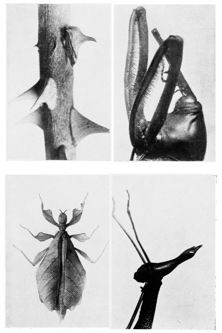
1. A Thorn Insect.—A striking example of protective resemblance. When resting on a thorny twig this little insect is safe from all its enemies. 2. The Head of Palm Weevil.—The long snout distinguishes the weavils from all other beetles. Its very long front legs are also worthy of notice. 3. A Leaf Insect.—Green in colour, this insect bears a remarkable similarity to a leaf. Its sluggish habits heighten the illusion. 4. The Head of stick Insect.—There are few more curious insects than the stick insects. The specimen illustrated has a very bird-like head.
Perch, pike, sole and some other fish have much more peculiar scales, known as ctenoid or combed for the reason that their unattached, that is to say their hinder margins, are toothed like a comb.
The scales of sharks, dog-fish and rays are called placoid for they are toothed; not only so but their arrangement is frequently quite dissimilar to the scales of ordinary fish. Taking the herring as our example, but a salmon or any other fish would serve equally well, and examining the arrangement of its scales with the help of our pocket lens, we shall find that the scales are fixed to the fish by their forward edges and that each scale partly overlaps its neighbour, as do tiles on a roof. In the shark family, however, the scales are often relatively wide apart, they do not overlap but are imbedded separately in the skin. The scales of rays have each a hard spine projecting from the centre, those of sharks and dog-fish have teeth, and they are teeth not only in appearance but also in structure.
The ganoid scales of sturgeon we are hardly likely to meet with. Sometimes these fish are on sale in London and other large towns and a specimen of their scales may be procured. They are bony in structure and, though interesting, require a considerable amount of preparation to render them sufficiently transparent to be examined under the microscope.
It is interesting to note that all the fossil fish[122] which are discovered from time to time have either ganoid or placoid scales, a fact which shows that the sharks, rays and sturgeon are directly descended from creatures which swam the seas thousands of years ago.
The shells of shell-fish are not easy to examine microscopically, but frequently their plates may be detached from the edges of such shells as oysters and mussels and these should be examined. If the outer part of the shell be taken we can easily see its honeycomb structure and, by adding a little acid and waiting till all action has ceased, we shall have a structure remaining which is remarkably like a number of plant cells. The inner layer of many of these shells is composed of beautifully iridescent mother-of-pearl. Now such iridescence is usually caused by surfaces furrowed with many very fine lines and mother-of-pearl is no exception. Under the microscope, with a moderately high magnification, we can see minute striations all practically parallel to one another.
The cuttlefish is peculiar in having a skeleton which is a moderately soft plate. These plates can often be found washed up by the tide, may be cut out from a dead cuttlefish or bought from a chemist’s as cuttlefish bone. However we secure the material we shall find that one side of the “bone” is hollow and that across this hollow, delicate plates run parallel to one another at intervals. Between these parallel plates there appear to be a number of fibres but, if we cut a thin slice of the structure and examine[123] it under the microscope, we shall see that the apparent fibres are really very thin plates of bone which wind and double upon themselves in a beautiful manner. The structure of these plates gives strength to the bone without adding to its weight.
Snails are sure to attract the microscopist sooner or later, so too are slugs. Many of the latter have shells, small flat or ear-shaped shells, quite different to the portable homes of snails. In many young snails, which may be killed by dropping into boiling water, we can find the shells so transparent that they form good objects for our microscope. Sometimes they are composed of six-sided cells, sometimes of beautiful star-shaped cells.
From the microscopist’s point of view the most interesting feature of the snail is its rasping organ, often wrongly termed the tongue. To find this organ it is necessary to open up the mouth of a dead snail, and if we seek the assistance of our lens while doing so, we shall have no difficulty in finding the rasp—it may be recognised by the minute teeth with which it is furnished. The whole structure should be carefully removed and mounted upon a slide. In some kinds of snails there are but a hundred teeth, other kinds, however, possess as many as twenty-six thousand eight hundred. The snail makes use of this remarkable organ to procure its food. Vegetation is pressed against a plate at the top of the creature’s mouth and literally filed into small pieces by the rasping organ. Captive[124] water snails may be watched while using their rasp upon the water plants or upon the green slime which soon accumulates in aquaria. The eggs of snails are easily found and should be examined, the queer little inmates may be studied through the transparent shells, in all stages of development.
The naturalist whose inclinations lead him towards the study of animal life will find plenty to occupy his time and his microscope. All kinds of eggs of small creatures may be watched as they develop. Frogs’ eggs are of interest in this respect, so too are tadpoles which hatch from them. The whole blood circulation in a young tadpole may easily be studied under the microscope, the structure of the external gills, the gradual change to internal gills, the development of legs, the absorption of the tail. The tadpole and its marvellous changes will afford sufficient microscopic material to last for many weeks.
The study of rocks and minerals by means of the microscope is apt to be disappointing. In the first place, to study them seriously we require a special microscope, the ordinary instrument, with which we may poke into the deepest secrets of the animal and plant world cannot translate for us half the story of the rocks. Again, to understand rocks and minerals we must study them somewhat deeply. Geology, as the science of rocks is called, is no more difficult than botany or zoology, the sciences of plants and animals respectively. Botany and zoology, however, appeal in some degree to nearly all of us; we may learn a good deal concerning the structure of the cockroach with the help of our microscope and be interested in the revelations of our instrument, but to embark on a detailed course of the minute internal anatomy of insects would appeal to few of us. Animal or plant life may be studied piecemeal and enjoyed on account of its absorbing interest. Geology must be studied from[126] its very beginnings if we are really to understand what we see beneath the microscope.
In the hand, a lump of rock, say of granite, may be of exceeding beauty. The body of the rock is, perhaps, a delicate pink, scattered here and there are the flat glittering plates of mica and brilliant crystals of quartz. Other rocks, less common, vie with the rare gems for beauty of colouring and lustre. As thin microscope sections these once gorgeous specimens are colourless, dull and, unless we understand them, uninteresting.
There are, however, many mineral substances which we may study with advantage for, if our investigations do not take us very far towards elucidating the story of the rocks, we shall at any rate discover something that is new to us. We may well commence our studies with the examination of ordinary sand. This is not a rock, you will probably exclaim. You are right but one day it may be a rock, it all depends upon circumstances.
Before we take out our microscope let us have a short talk about rocks in general, then we may understand better where we are. Rocks of one kind and another make up the crust of the earth, that is pretty obvious anyway. Thousands and thousands of years ago, how many we are not prepared to guess, this old earth of ours was a sphere of molten rock. Needless to say it was far too hot for any plants or animals to dwell upon it. Very, very gradually the outer crust cooled down and in time it became sufficiently cool to support animal[127] and vegetable life. Then there were rivers and seas and then came, from time to time, rain and wind and frost and even earthquakes. The earthquakes cracked the crust of the earth, moisture entered the cracks and, when the frosts came, pieces of rock were broken away, owing to the expansion of the moisture in the cracks as it became converted into ice. The rain and wind helped to carry the broken pieces of rock, ever downwards towards the sea, but before the sea was reached the big boulders became broken up, by their buffeting, into shingle and sand and mud. In the course of long ages, longer than it is easy to imagine, these broken pieces of rock, gathered together as we have seen from various districts, may have been left high and dry, for the face of the earth has not always been as we know it now.
In time all the little particles became welded together to form a new kind of rock. Sometimes animals and plants were buried in the mud destined to become a rock and their parts were so well preserved that they may not only be recognised by present-day scientists, but in many cases their structure may be made out so well with the aid of a microscope that no one would guess they had been buried thousands of years ago.
New rocks have been formed not only by the breaking up and welding together of the original earth’s crust, but by animal and plant remains. In some places, in past ages, billions and billions of little shell-fish have lived in the waters, died there[128] and their shells have fallen to the bottom of their watery home. Now we know their shell remains as chalk or if it has undergone great pressure, owing to changes in the earth’s surface, as limestone. There are rock masses also formed of the remains of countless Diatoms; sponge spicules, too, have played their part in rock formation.
It is clear from what we have written that, in the first place, the rocks of the earth form two great divisions, the rocks which formed the original crust of the earth, Primary rocks they are called, and the later-formed Secondary rocks. The Primary rocks are often glassy in appearance, they show unmistakable signs of having once been molten and they never contain animal or plant remains, fossils as they are called, for the reason we have already explained. The secondary rocks often betray their origin by occurring in layers, or strata to speak more scientifically. Always when we think of the rocks we must think in thousands of years, then it will be easier to understand the formation of these strata, each one of which may represent the work of hundreds and hundreds of years.
Now to return to our sand; we have explained briefly that sand is really the remains of broken up rocks. First we have the solid rock, boulders are broken from it as the boulders are acted upon by rain, frost and wind: they become more and more broken up and at the same time they are carried towards the sea where they take the form of shingle. The shingle, in the course of time, becomes broken up into sand, and the sand again becomes so finely divided that it forms mud. From this it is clear that the composition of the sand depends largely on the nature of the rocks in the neighbourhood.
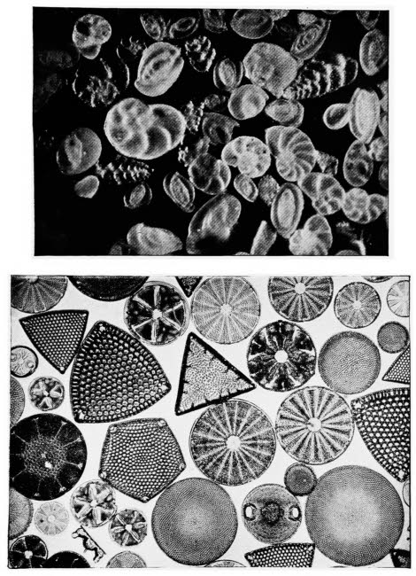
By the courtesy of Messrs. F. Davidson & Co.
1. Foraminifera
Small sea shells. The inmates die and the shells, falling to the bottom of the sea, are gradually converted into chalk.
2. Diatoms
Small water plants which make beautiful objects for the microscope by reason of their remarkable shapes and sculpturing.
First of all, we must place a little of our sand upon a piece of black paper and examine by reflected light. We shall soon notice that sand is anything but the simple gritty looking substance it appears on the sea shore. At least four different substances are certain to be seen; if the sand be clean, we shall see bright glassy crystals of quartz; angular, sharp-edged pieces of flint; minute, flat, glistening plates of mica and the broken remains of shells. Should our sand be taken from a spot in the vicinity of volcanic rocks, it will probably contain opaque pieces of magnetite; we may not recognise these when we see them, but they may be separated from the rest of the sand by means of a magnet, to which they will adhere. Probably our sample of sand will be dirty or stained with iron, in this event we may wash it with weak acid, then after drying, we may examine by transmitted light and the various crystals will show up well.
Let us take some clay as our next example. Clay is the substance from which the rocks known as shale and slate have been formed. We cannot see much of the structure of clay with our microscope so let us wash it and, by so doing separate it into its components. We must put a little clay into a tumbler of water and stir it vigorously for[130] some little time, then still stirring, if we pour off the muddy liquid into another tumbler, we shall find that our old friend sand has settled to the bottom. Clay then is merely mud and sand, but we must not throw this sand away without examining it for, very frequently, beautiful minute fossils are to be found amongst it. Little creatures dwelling in the mud, become buried in it and, as more and more mud is formed above them, and it becomes partly solidified in the form of clay, they become fossilized.
If now we examine a piece of sandstone with our pocket lens we shall find that it very closely resembles the sand we have already studied. It may be so soft that we can break it up in our fingers, then if we examine the powdered sandstone beneath the microscope we shall need to be experts to tell whether we are examining sand or sandstone. Should our rock be too hard to break up in our fingers the addition of a little weak acid will reduce it to sand. We see now that a large proportion of sand and a small proportion of mud result in the formation of sandstone, whereas if the proportions are reversed and we have more mud than sand the resulting rock is known as shale or slate. In shale we very frequently find fossils, in sandstone rarely, for the reason that structures buried in the mud, destined to become shale, are protected from the atmosphere and the action of water; sandstone, on the other hand, is porous, moisture trickles through it and any delicate structures which may have been buried in[131] it are more likely to decompose than to become fossilized.
Limestone is easy to obtain and may occupy us for a few moments. We do not wish to go into technical details, but we may say that the word limestone includes a number of rocks which differ largely in appearance and to a considerable extent in composition. It would, perhaps, be more correct to say that there are several kinds of limestone. Some kinds are made up almost entirely of shells and very interesting they are as microscopic objects. One may wonder how a geologist can state with certainty that some spot, may be many miles from the sea, was once covered by salt water. One of these shell-formed limestones may give him the information; he knows that certain shells comprising the rock must have belonged to marine animals, he knows too that whole mountains, which these rocks sometimes form, are not carried bodily on to dry land, so the obvious inference is that the rock was formed below the sea.
The softer limestones may easily be crumbled and powdered for examination under the microscope; the harder kinds should be treated with acid. When acid is added to limestone a considerable effervescence takes place, for the acid decomposes a substance known as calcium carbonate, which the limestone contains, and bubbles of gas are given off. When the limestone has ceased to effervesce the portions of the rock which remain, may be carefully washed in water, dried and examined under the[132] microscope. We shall find that the substance we examine consists of sand containing a goodly number of plates of mica and there may also be a number of sponge spicules. We must bear in mind when we are examining the remnants of limestone after treatment with acid, that shells are largely composed of calcium carbonate, so it is useless to look for any shell remains, for the acid will have dissolved them.
Calcium carbonate, in a nearly pure state, will and does form rock-like structures; we are most of us familiar with stalactites which are formed on the roofs of caves. These structures are usually composed of calcium carbonate though sometimes, notably in some of the Derbyshire caves, Barium takes the place of Calcium. Should we have the chance of examining a section of a stalactite we should certainly do so. We can see the rings which are formed, as layer after layer of calcium carbonate is deposited, in fact the section of a stalactite bears a striking resemblance to a stem of a tree and has, before now, been mistaken for a fossil stem.
If the study of rocks appeals to us we should make a point of examining all the specimens we can lay hands upon. Many quite common specimens may easily be obtained; rock-salt, for example, though in itself not of great interest as an object for the microscope, will readily dissolve in water, leaving behind an insoluble residue of iron which is well worth examination. The majority of rocks, however, are not affected by water and but[133] little by acids. With such specimens the only course open to the microscopist is to prepare sections.
The making of rock sections is certainly different to the cutting of plant or animal sections. It is a laborious business as the enthusiast will find to his cost. The professional makers of rock sections have special grindstones or lathes for the purpose and, even so, the process is not rapid. The amateur must needs do all his preparation by hand. The requirements, in addition to the piece of rock of which we require a section, are a small square of plate glass, some Canada Balsam, emery powder of various grades and an unlimited stock of patience. If possible we choose a piece of rock with one side as nearly as possible flat; this is merely to save labour; the piece of rock should be roughly about half-an-inch square. As a start we rub the flattest side in a mixture, practically a paste, of coarse emery powder and water. As a matter of fact, we may keep the piece of rock in our pocket and grind it when occasion offers on a flat stone wall or on any surface that will assist in producing a flat surface. When we have ground our surface flat and smooth, we finish it off with fine emery powder and may then polish it with jeweller’s rouge. So much for the first side and, if we do not cry enough at this period, we may proceed to the grinding of the other side. Taking our slab of plate glass, we fasten the polished side of our piece of rock to it by means of Canada Balsam, then we may rest for a few days while the Balsam sets. As soon as the[134] rock is firmly fixed to the glass we proceed as before, but in this case the final grinding and polishing must be very carefully carried out. Towards the end of the operation our original lump of rock will be reduced to the thinness of a cover slip and to its fragility. Having given the finishing touches with jeweller’s rouge, we put the glass, with its attached rock section into a bottle of xylol (to be obtained from any chemist). The xylol dissolves the Canada Balsam and the rock section falls from its support. A further washing in clean xylol should be given and then the section is ready for mounting, which is best done in Canada Balsam. The section may be carefully fixed to the slide with a drop of Balsam or it may be covered with a cover slip, in the usual manner.
In our concluding chapter we give hints on slide making and addresses of firms who supply slides. Our advice in that chapter is to prepare one’s own slides where possible; in the case of rock sections, however, we must change our advice, only the microscopist of unlimited leisure can find time to make his own slides.
One of the most interesting branches of rock study deals with the fossil remains of plants and animals. Fossils are interesting in themselves: they are doubly interesting because they tell us more than we could ever have discovered without them, concerning the living forms which inhabited the earth at different periods. Geologists know the order in which the various secondary rocks were formed and by studying[135] the fossils of these various rock formations, from the earliest to the latest, can tell which animals and plants have been longest upon the earth. Some of the living forms of to-day have existed from very early times; we know, for example, that cockroaches were upon the earth long ages ago, for their fossil remains are found in the very early rocks.
For the most part the fossils of animal forms do not make good objects for the microscope. The Foraminifera, minute creatures dwelling in shells, are the most suitable for microscopic examination and very beautiful some of them are. They are best examined by reflected light, for then their little shells show their delicate pearly sheen.
Sponge spicules, as may be guessed from their hardness, are common in the fossil state; there are also fossils of sea-anemone ancestors and fossils also of Hydra colonies. The last named, known as graptolites, look not unlike lead-pencil marks on the rock. They exist in many forms; Diplograptus has a stem with two rows of cups in which the little creatures lived long ages ago; Monograptus has but one row of cups upon its stem, whilst Didymograptus is a branched form. These fossils are by no means rare and are worth studying under the microscope.
There are curious fossils knows as Trilobites, not unlike the present-day wood lice; they too may be studied, for some of them show all their characteristic markings as plainly as they must have appeared on the living animals.
In the plant world, many fossils in amazingly, good states of preservation have been found. Some of the giant Club Mosses from the coal measures, exhibit all the characteristic stem markings, leaf scars and the like, so clearly that one might imagine one examined a living specimen. Most of the plant remains are beyond the reach of the amateur, but many of them may be viewed in museums, in different parts of the country and the microscopist, whether he be a student of rocks or a lover of plants, should make a point of examining them. From quite fragmentary remains, scientists have been able to conjecture what vegetation covered the earth at various ages. The present is the age of flowering plants, but long ago the world was green with giant Club Mosses and Horsetails, very humble plants at the present time.
It is an unfortunate fact that our food is not always absolutely pure. It may be contaminated with foreign matter either by accident or by design. However careful the manufacturer may be in, say the preparation of cocoa, some dust, some waste vegetable matter, perhaps even a few stray dried insects may occur as impurities. They are out of place certainly but, at the worst, they are a sign of lack of care on the part of the manufacturer. There is another, more serious side to the question of food adulteration, where the foreign matter is added purposely, either because it is cheap, because it weighs heavily, imparts a pleasing colour or an agreeable aroma. Such adulteration is a form of fraud and the microscope is an invaluable aid in its detection.
In many respects the microscope is a better informant than the tests of the chemists; in some cases, however, it merely supplements and confirms the chemical results. Let us consider, for a moment,[138] the advantages possessed by the microscope and also where chemistry scores.
Very frequently the results of costly law cases hang on the reports of expert food examiners; every care, therefore, must be taken to avoid error. This being the case, whenever possible, chemical tests should be carried out to confirm the results of microscopic examination. When both microscopist and chemist come to the same conclusion, there is not likely to be any mistake. There are tests which the microscope cannot perform, there are some, also, which are beyond the powers of the chemist and many which are very difficult for him. A drop of milk, for example, examined under the microscope shows a number of fat globules floating in a watery liquid. However clever the microscopist and however accurate his instrument, he cannot tell if there is an excessive quantity of water, yet a simple chemical test will answer the question. This is a case in which the microscope is of little use, although it is only fair to add that microscopic examination would reveal the presence of blood, hair and dirt, to mention three common impurities, which the chemist in his test for watered milk would quite overlook. With a little care and the use of suitable stains, any bacteria which might be present would also show plainly under a powerful microscope.
Now for an example or two where the microscopist has the advantage of the chemist. Some jam makers have been known to be sufficiently unscrupulous[139] to sell “raspberry” jam contaminated with a large percentage of some cheaper fruit, such as gooseberry. The seeds of the two fruits differ so markedly that it is really not necessary to employ a microscope to discover the fraud, but a case is on record where wooden seeds were used, so like the true seeds of the raspberry, that a very careful examination was necessary to show what had happened. In our chapter on the Microscope in Agriculture we have referred to this point in greater detail. Starch of various kinds is a very common food adulterant and the experienced microscopist can estimate almost precisely, the proportions of different starches in a mixture, a feat which would sorely puzzle the chemist. So in certain cases the microscope is indispensable.
Briefly the microscope is a time saver; chemical tests occupy a considerable time; microscopic examination is quick, the experienced microscopist at once recognises what he observes. Very small quantities can be examined under the microscope, relatively large quantities are required for chemical tests. Again, if only a small quantity of the material is available for examination and it is necessary to carry out chemical tests, they can be performed under the microscope and this point is considered in another chapter.
We have mentioned that starch of various kinds is a common adulterant of many foods and the budding food analyst might do worse than learn to recognise the various starch grains under the[140] microscope. They are easily obtained and as easily observed. Each variety of starch has grains which are remarkably constant in their characteristics. A beginning may profitably be made with potato starch, for its grains are large and they possess certain well-marked features, which may or may not be present in the grains of other starches. By scraping the newly cut surface of a potato we can obtain thousands of starch grains. The surface of the potato must not be grated, just a gentle scraping with a pocket knife and a mere speck of the cloudy liquid that is obtained, added to a drop of clean water on our slide, will suffice. Cover the object with a cover glass and examine under a fairly high magnification. There are countless, oval, almost transparent bodies in our field of view, they are potato starch grains. Each one, as we shall see when we make a more careful examination, is not unlike a miniature oyster-shell. In the shell, there is a point which is its oldest part and the remainder has grown, layer by layer, round that point till the shell is fully formed. Now we magnify the starch grains as highly as possible and slowly rotate the fine adjustment to and fro, for the reason that the object is not flat and by doing so, we obtain all its parts in focus in turn. If the illumination is not too intense, we shall notice a minute dark dot corresponding to the oldest part of the oyster shell; it is, in fact, the oldest part of the starch grain. Around this point we can see as we focus up and down, ring after ring where the grain has grown[141] larger and larger. The dark spot is called the hilum and the rings are known as striations. In the potato starch grain the hilum is not central and the striations are not circular. Wheat has large and almost round grains without a hilum or striations, those of Barley are very similar but smaller and not so uniformly round. Rye grains are frequently cracked and often have ragged edges.
A very large number of these objects may be examined, for it is useful to know their structure if one’s object be to examine various foods; from the point of view of beauty, when examined with a polariscope, they have few rivals. Maize starch, which is to be found in most houses under the name of corn flour consists of two kinds of grain. Some are many sided and angular, all of one size and without striations, they are also split at the centre; the other grains are rounded, of various sizes and are never like the angular grains grouped together. The former come from the horny part of the maize, the latter from the floury portion.
Rice starch is also many sided and angular, almost crystal like; there are, however, never any rounded forms and this serves to distinguish it from maize starch. The shape of Arrowroot starch grains varies according to the plant from which it is derived, for this substance does not all come from one kind of plant but, whether the grains be pear shaped, hammer shaped, triangular or dumbell shaped they all show striations and an x-shaped split in place of a hilum. Tapioca starch grains are[142] usually grouped together in twos or threes; when they rest on their flat surfaces they appear circular and each hilum is surrounded by a dark ring, when on their sides they are seen to be sugar-loaf shaped.
Many more starches can be found without going far afield, Sago, Peas, Beans, Lentils and Bananas are a few common commodities containing starch. An effort should be made to study the very curious dumbell shaped starch grains of the Spurge and its relations. All these plants contain a white milky juice in which the starch grains float; by squeezing a little of this milky fluid into a drop of water on a clean slide the grains can easily be observed.
It is sometimes difficult to observe starch grains till a fair amount of experience has been gained in the use of the microscope. Should this difficulty arise, it may be overcome by adding a drop of a weak solution of iodine. This will stain the starch grains a deep blue colour and render them very easy of observation. The iodine solution must be weak, however, or the staining will be excessive and the objects rendered black and non-transparent.
Having examined many or all of the specimens we have mentioned let us turn our attention to some of the common foods, and learn some of the methods used in testing for impurities. Ordinary household bread, it is hardly necessary to state, is rich in starch and, by trying the iodine test, mentioned above its presence is easily shown. With a weak solution the deeper the blue colour produced, the[143] greater the quantity of starch. Some parts of the bread will be stained yellow, this indicates the presence of another nourishing component of bread. Certain kinds of bread are supposed to contain no free starch, because this substance is not beneficial to some people. Iodine again will reveal whether the bread is as it is described, for, if there be no free starch there will be no blue colouration. Brown bread will show much more of the yellow colouration and less of the blue than white bread. Good, well-baked bread should keep for a considerable period without turning sour; we can easily see whether our sample is satisfactory by running a drop of litmus on to it and watching the effect under the microscope; if the litmus remains unchanged in colour the sample is not sour; if, on the other hand, the litmus turns red it shows us that acid is present and that our bread is not as it should be.
Tea is difficult to prepare for microscopic examination and most of the tests call for expert knowledge, not only in the management of the microscope but of the plant itself. The structure of the leaves can be made out clearly in specimens which have been soaked for a time in water, but this is of little interest to the ordinary microscopist. One very pretty test may, however, easily be performed. We all know that it is not good to drink tea which has been standing for a long time. Some tea-drinkers are so particular that they cannot bear to see the teapot shaken before they have poured out their cup. All[144] this trouble arises because tea contains a poison called “theine”; it is an alkaloid, one of a large class of chemical substances which are nearly all deadly poisons—cocaine and nicotine are alkaloids. Although theine is poisonous, tea which contained none of this substance would be tasteless and the absence of this substance shows that the tea leaves have been badly prepared. Tea after being gathered should be dried at once, sometimes it is re-dried and this process drives off the theine. For our test we require, in addition to our microscope, two watch glasses, a piece of copper wire gauze and a gas burner or a spirit lamp. Place a little tea in one of the watch glasses and cover with the other watch glass; then heat gently on the wire gauze. In a few minutes drops of moisture will appear on the upper watch glass; after about ten minutes’ heating beautiful, long, needle-shaped crystals will begin to appear, with a little further heating we shall obtain a good crop of lovely crystals on the upper watch glass and they make a splendid object for examination under a low magnification. The crystals are of theine, the poisonous component of tea, and the test is used to discover whether the tea has been redried during its preparation; redried tea gives no crystals.
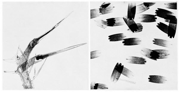
Photos by Flatters & Garnett
The Stinging Hairs of a Nettle
These hairs are much longer than ordinary plant hairs. Sharply pointed at one end, there are sacs at their bases containing acid.
Butterfly Wing Scales
Scales from the wing of a butterfly. Each scale is a hollow sac, affixed by its notched end to a pit in the insect’s wing.
The examination of cocoa for impurities is a matter rather for the chemist than for the microscopist. It contains a vast number of starch grains, not unlike those of rice, except that they are rounded. Coffee often contains a number of impurities,[145] the chief being chicory, various starches, ground acorns and date stones. Chicory is really an impurity, though it is one often asked for by coffee-drinkers. It is easy to detect the amount of chicory present in a sample of ground coffee, by throwing a little of the mixture on to water. The chicory sinks at once, whereas the coffee floats for a while because it is oily. In pure coffee there should be no starch and the iodine test will readily show whether we are dealing with a sample free from starch or not.
Mustard is very rarely purposely mixed with any impurities, in fact it is probably the least likely to be adulterated of any article of food. Under the microscope a large number of small objects, very similar to starch grains, can be seen. They are the cells containing mustard oil and they are not stained blue by iodine. A specimen of pure mustard contains no starch. Pepper is by no means easy to test for impurities. It contains minute starch grains, which can be recognised under the microscope after staining. It is mentioned here because of a very interesting and easily performed experiment that will appeal to every microscopist. Place a little pepper on a clean slide and moisten it with a drop of alcohol, allow it to stand for a minute or so then add a little dilute glycerine, cover the specimen with a cover glass and examine it under the microscope after the lapse of about five minutes. The sight of wonderful prismatic crystals forming one by one in rapid succession will be ample[146] reward for the trouble taken. A drop of strong nitric acid, which must not be allowed to come in contact with any part of the microscope or with one’s hands or clothes, will turn the crystals a rich orange colour. The crystals are composed of a substance called piperine.
Everyone knows of the importance of pure water for drinking purposes but the word pure in this case is used in a very wide sense, for the only really pure water is distilled water and it would not form a very good beverage. Although ordinary tap water may contain a number of impurities it is not easy to see them without taking a little trouble. If our tap water is so contaminated that a drop, examined at random, shows us all manner of solid matter floating in the water there must be something seriously wrong. Those who are engaged in testing water under the microscope, first take a big jug full of the water and allow it to stand for twenty-four hours, covering it the while to keep out dust. At the end of this time most of the water is carefully drawn off, from the top, with a siphon and the remainder, after stirring is poured into a conical glass and allowed to stand for a further twelve hours. Then the upper portion of the water is again siphoned off and a little of the remainder, which is left in the point of the conical glass is drawn by suction into a special kind of glass tube, called a pipette. This final sample contains all the solid matter which settles to the bottom of the water after standing for thirty-six hours.
The impurities likely to be present in water make such a formidable list that we can only mention a small number. There may be various mineral substances, such as lime, sand or clay; vegetable substances, starch grains, fragments of wood, pieces of decayed leaves and the like or there may be hair, scales of fish, etc. The impurities may be living plants, of which the most likely to be found are bacteria, diatoms, desmids and Volvox, amongst plants, and rotifers, Vorticella, Paramœcium, Amœba, also portions of tapeworms and their eggs amongst animals. These creatures are all described elsewhere so we need not dwell on their peculiarities here.
In addition to all of the above and many more not mentioned there are four metals commonly found in impure water, either in small solid particles, or in the form of one of their compounds soluble in water. The metals are iron, copper, zinc and lead. Very simple chemical tests will show whether they are present or not. To a drop of the water add a very minute quantity of hydrochloric acid and of potassium ferrocyanide solution. When iron is present the solution will turn blue; in the presence of copper it will become bronze coloured, whilst zinc turns it a milky white. To detect lead, take another drop of water and add a very small quantity of potassium chromate solution. If the suspected impurity is present the solution turns yellow. All the chemicals for these simple tests may be obtained at any chemists and there is this great advantage[148] in testing under the microscope—only very small quantities are required.
Butter can hardly be described as an interesting object for the microscope, nevertheless, it may be of use to explain the methods of its examination. A small sample should be placed upon a clean slide, a drop of olive oil added and the whole covered with a cover slip which may be pressed firmly till the specimen forms but a thin layer. Of course the most important impurities likely to be present are bacteria but these we cannot see without special preparation and we are not dealing with bacteria in the present chapter.
If our specimen is in a film sufficiently thin to be transparent, and we should have made it so by pressure on the cover slip, we may first of all examine it carefully for starch grains which, by the way, should be absent. The amount and size of the drops of water, which every butter sample contains, are of importance in deciding its quality. In good butter there are a few scattered drops of various sizes; in milk-blended butter the water globules are all very small and all of the same size, or as nearly so as the eye can judge; in butter containing an excessive amount of water the drops appear large, much larger than in a normal sample.
If we examine various samples, we shall find that some are much more transparent than others, the transparent samples being fresh butter. The curd also in fresh samples is much more finely and evenly scattered in the field of view than in older samples.[149] Renovated butter, that is to say rancid butter which has been melted and made palatable by forcing steam through it, should be examined by oblique light—easily arranged by tilting the mirror at an angle—when the curd appears as white patches on a dark background.
It is curious that one article of food, honey, is more likely to be pure when it contains impurities. This sounds like a bull but a great deal of honey is manufactured from various sugars but not by bees. This artificial honey contains no pollen grains, in fact any honey found to be free of pollen should be looked upon with suspicion. Starch often occurs in artificial honey, never in real bee-made honey.
To many foods adulterants are added as preservatives, the nature and quantity of such additions is settled by Act of Parliament. Many foods are preserved with small quantities of Borax or Boric Acid. The use of Formaldehyde, formerly sold under the German trade name of Formalin, is not unknown but it is very injurious. Salicylic Acid which was formerly much used is being supplanted by Benzoic Acid, for the reason that the latter is not so easily detected and therefore prosecution for excessive quantities is not so likely to follow. These preservatives are not easily detected by the microscopist unless he be a chemist also.
As we have already remarked adulterants are added for the sake of colour, either because the public demand certain colours, or to hide fraud. Milk for instance, when watered, assumes a characteristic[150] blue colour; to hide the blue shade various dyes, anetto, turmeric or carrot juice are added or one of the aniline dyes, products of coal tar. This form of deception became so common that now the public demand yellow milk and butter. Jams made from inferior or over-ripe fruit are frequently coloured with coal tar dyes, so also are cheap sweets. Oxide of iron is added to potted meats, sauces, etc., to improve their appearance. Bottled peas, which if untreated would be of a yellowish-green colour, are made to appear bright green by the addition of the poisonous blue vitriol; fortunately this chemical unites with the chlorophyll of the peas to form a compound which is insoluble in the human body and so no great harm is done. We may compare the chlorophyll in a healthy, undoctored green pea with that in a pea which has been treated with blue vitriol; under the microscope we shall notice the striking difference between the two.
When we admire the beautiful crystals which go to the making of a piece of lump sugar we little dream that, if those crystals were pure they would be yellow. The housewife, however, demands her lump sugar white so ultramarine is added to mask the yellow colour and give the sugar its white appearance.
Most of these impurities are difficult or impossible of detection under the microscope; they are added to give the food a more pleasing appearance in the first place, for there are undoubtedly certain people who prefer to consume food which appeals to the[151] eye, though of doubtful purity, rather than unadulterated though perfectly pure fare. When added solely for the sake of appearance it matters little, but the habit of making these additions is frequently cultivated to hide bad material and imperfections in manufacture.
Sometimes adulterants find their way by accident into our food. A good many years ago numbers of people were poisoned by drinking beer, in some cases with fatal results. Tests were made and the beer was found to contain arsenic but how it got there remained a mystery. At length the glucose, a kind of sugar used in making beer and added also to a good many of our foods, was found to contain the substance. Now in the making of glucose, sulphuric acid is used and in this particular case the impure or commercial acid had been taken. This impure acid frequently contains arsenic and, in the case we mention, the results of its use were disastrous.
There is probably no scientific work more wedded to the microscope than the study of bacteria. We may learn a great deal about birds or insects or rocks or minerals, without any instrument but we can learn little of the bacteria unless they are highly magnified.
There is such an extraordinary amount of misconception concerning bacteria that, it will be time well spent if we attempt to clear up all misunderstanding at the start. Bacteria, often called microbes or germs, are looked upon with considerable awe by most people, who associate them in some vague way with disease. There is no denying that many bacteria are responsible for certain diseases; many more are perfectly harmless and a goodly number are exceedingly useful.
To enumerate all the bacterial activities would require a large book but briefly, apart from the disease-causing bacteria, they enter into the manufacture of cheese and butter, of wine and vinegar;[153] they are essential to brewing and tanning; they act as scavengers over the face of the earth, breaking up a mass of decaying animal and vegetable matter into simple chemical substances which can then be used again as food for plants; some of them also can take a gas called nitrogen, from the air, and pass it on to green plants.
What are these active little creatures? The question is a natural one. They are merely very minute, one-celled plants. Each one possesses a firm cell-wall, filled with living matter; in an earlier chapter, we described the one called protean animalcule and, although it was composed of but a single cell it had no definite wall. This is one of the essential differences between plants and animals, both of them are made up of one or more, maybe millions of cells, but each plant cell is surrounded by a well-defined wall, animal cells have no such walls. The exact position of the bacteria in the plant world is still open to doubt. Most scientists place them amongst the fungi; for, with very few exceptions they possess no chlorophyll. One or two of them, however, do possess a green colouring matter which, if not chlorophyll is very near to it; on this account other scientists are of the opinion that they are related to the seaweeds. It is a matter, however, that does not concern us very deeply, for our purpose it is sufficient to know that they are plants. When they were discovered, nearly three hundred and twenty-five years ago, they were looked upon as minute animals and it is curious[154] that the belief has survived this long period of time in the popular mind. Long before the activities of bacteria were connected with various phenomena, such as infectious diseases, souring of milk, etc., it was thought that these changes were brought about by chemical action. Like many of the early theories, this one contained a half truth, for a great many of the changes brought about by bacteria are really due to chemical action initiated by the organisms. In other words, the bacteria set free certain substances which actually cause the changes to take place.
Let us make our statement clear by a simple experiment. To a little fresh milk we add a weak acid, the milk curdles at once and by dipping a piece of litmus paper (obtained at any chemist’s) into the mixture, it will turn red, showing the presence of acid. Litmus, by the way, is obtained from a lichen; in the presence of acid it is red, an alkali, the opposite of an acid, turns it blue. In a neutral solution, that is to say one that is neither acid nor alkaline, litmus is of a purplish hue.
To continue our experiment, we allow another sample of the same milk to stand for a day or two in a warm, dark place and again the milk will be curdled. A test with the litmus will show that the solution is acid. The bacteria themselves have not curdled the milk but they have liberated a substance, called a ferment, which has split up part of the milk into an acid, amongst other things and that acid has actually done the curdling. For this[155] reason, weak alkalies are sometimes added to milk. Acids and alkalies, of equal strength form neutral solutions, so that, when the milk bacteria begin their activities which result in the formation of acid, it is at once made neutral by the alkali. By this means, curdling is postponed for a little while, though there comes a time, of course, when all the alkali is used up, then the acid gains the upper hand and curdling takes place. We could if we wished continue adding more and more alkali to keep pace with the formation of acid, but too much alkali would be as unpalatable as too much acid, so nothing would be gained.
Before we bring out our microscope to examine these lowly plants, we will first of all kill a myth which has survived, in the non-scientific mind, since the eighteenth century and then describe briefly the life history of a typical bacterium. Now for the myth. Bacteria are so minute, their activities so great and the results of their activities so far reaching, that it is hardly surprising to learn that, even at the present day, bacteria are supposed simply “to happen.” We talk of thunder turning milk sour, but thunder can no more sour milk than can a passing train or an air raid or a burst in a water main. True, milk turns sour more quickly in thundery weather than in frosty weather, because, when thunder threatens, the air is warm and the milk-souring bacteria increase more rapidly in warm weather than in cold. We must remember always that bacteria are living beings and in common with[156] all other living things they must have parents. What probably took place at the beginning of the world we cannot consider here but one thing is certain that, at the present day, no living matter is produced from non-living matter; “life from life” is the only theory that will stand scientific tests and this has been the case ever since the simplest microscopes were thought of and thousands of years before that. Any substance, however liable to decay, if rendered germ free and kept germ free, will retain its fresh condition indefinitely. Could bacteria or germs, call them what you will, simply happen it would be useless attempting to fight against them.
Bacteria are everywhere. In the water we drink, in the milk, butter, cheese and in dust. We cannot avoid them, try as we will; it is fortunate, therefore, that the majority are harmless. You may be surprised that, with this ubiquity, you have never seen one. When, however, you learn that most of them are only about twenty-five thousandths of an inch long and that a thousand million of them could be packed comfortably into a little box, whose sides measured but a twenty-fifth of an inch in length, it is not really so surprising after all. Being so small, the activities of a single bacterium are insignificant; that “union is strength” was never better exemplified than amongst these lowly plants. There are no male and female bacteria, in the majority of cases they increase by splitting, in fact they are often called splitting plants. The change[157] may be watched under the microscope. The plant elongates somewhat, it becomes narrower and narrower in the middle, it develops a waist in fact; finally the two halves part company and each one leads a separate existence as a bacterium. This splitting progresses at an extraordinary rate. A celebrated scientist once wrote: “Let us assume that a microbe divides into two within an hour, these two into four in the next hour, these again into eight in the third hour and so on. The number of microbes thus produced in 24 hours would exceed 16 1/2 millions; in two days they would increase to 47 trillions, and in a week the number expressing them would be made up of 51 figures. At the end of a day (24 hours) the microbes descended from a single individual would occupy one fortieth of a hollow cube with edges one twenty-fifth of an inch long, but at the end of the following day would fill a space of twenty-seven cubic inches, and in less than five days their volume would equal that of the ocean.” It is hardly necessary to add that these alarming figures represent what would happen if no accident befell the bacteria, they show the enormous vitality possessed by the smallest of all plants. Even allowing for misadventure their increase is alarming; actual tests, with a sample of milk containing originally 153,000 bacteria per cubic inch, show that the cubic inch contained after one hour, 539,750; after two hours, 616,250; after seven hours, 1,020,000; after nine hours 2,040,000 and after 25 hours 85,000,000 individuals.
The writer whom we have just quoted calculated that a single bacterium weighs about 0.000,000,000,024,243,672 of a grain, that forty thousand millions weigh one grain and that two hundred and eighty-nine billions weigh a pound. The descendants of one bacterium weigh 1/2666 of a grain, after twenty-four hours; more than a pound after two days, and sixteen and a half million pounds after three days. The assumption in this case, also, is that no harm comes to any of them; the mortality amongst bacteria is, clearly, very great.
Sometimes, owing to external conditions, such as lack of food certain bacteria produce spores. The power of spore formation is not possessed by all bacteria and those which are able to bring it about are difficult to kill for the spores, which contain the living material of the bacterium are surrounded with walls which will resist boiling, drying, freezing and all manner of ill treatment. The spore formation of bacteria is very simple, all or part of the living contents of the bacterium becomes surrounded by a tough wall and remains so surrounded till circumstances are favourable, when the wall bursts, its contents escapes and becomes a bacterium, capable of founding a new colony by the method of splitting we have already described.
Now let us try to find out what sort of plants we are to look for, when we are searching for bacteria, under our microscope. They exist in many forms, to which special names have been applied, and it is unfortunate that, very often, their external[159] form varies according to their state, thus a bacterium may be spherical when young and rod shaped when older. Some bacteria are spherical and are known as Cocci or Micrococci, from Greek words meaning a berry or a little berry respectively; sometimes these spherical bacteria occur in pairs, then they are called Diplococci (double berries); or in chains, Streptococci (chain berries); or in bunches, Staphylococci (grape berries). They may resemble short rods, when they are called Bacteria, a name, by the way, which is also applied generally to all microbes; they may, on the other hand, have the appearance of longer rods and then they are called Bacilli. Some of these longer rods may be curved or even corkscrew shaped when they are known by the name of Spirilla. Rather fearsome names some of these we fear and we wished to avoid long names, but they appear over and over again in books and papers relating to bacteria so we are compelled to introduce them to our pages. Many bacteria possess no power of movement, others swim rapidly, by the aid of the lashing movement of little whip-like structures with which they are furnished.
After all this preamble, which we hope has cleared up certain misconceptions regarding bacteria and has given the reader some insight into their habits, we may proceed to the examination of some of the plants themselves. At the outset we have a confession to make. Bacteria can only be studied seriously, by those who possess very expensive and[160] elaborate apparatus; considerable technical skill is required to prepare the plants for examination—many of them indeed can only be seen after they have been stained and lastly, to trifle with the disease-causing members of the family may lead to dangerous if not fatal results.
Having issued our warning let us see what we can do in the way of microscopic investigation. The easiest subject with which to make a start is the Hay Bacillus, Bacillus Subtilis, not because it is the largest of the bacteria by any means, but because it is very easily obtained. Each plant measures about five thousandths of an inch in length, so we shall require a high magnification to examine it. Having obtained a small quantity of hay, we must boil it in water for about three-quarters of an hour and then set it aside for some hours. In due course the water will contain hundreds upon hundreds of bacteria or, speaking more correctly, of bacilli. For our work, we shall require a special kind of microscope slide; instead of the piece of plain glass we have been accustomed to use we must obtain one with a circular portion, hollowed out from the centre. Having done so, we take a clean glass rod and, with it, transfer a drop of the water, containing the bacilli, to the centre of a clean coverslip. Invert the coverslip so that the drop is on the lower surface and place it over the hollow portion of the slide, in such a manner that the drop still remains suspended from the coverslip; this is known as the hanging-drop method and requires[161] some little skill to accomplish satisfactorily. When our slide is prepared, with a magnification of at least one thousand diameters, we may reasonably hope that our trouble will be rewarded.
At first we shall probably see nothing. We recall that we had some difficulty in examining starch grains, on account of the fact that they were colourless. This time we are dealing with a far more difficult subject. When our eyes become accustomed to the light, however, we shall be conscious that there is something moving in our drop of water. The Hay Bacillus is one of the moving forms, each individual is furnished with a number of little whips whose lashings enable it to travel through the water. The whips cannot be seen in unstained bacilli; experience, however, tells us that they are there, for all these lowly plants which show movement are seen when stained, to possess the little whips. The process of staining kills the plants so that we cannot see the little whips in action.
Having detected that movement is taking place, a little adjustment of focus and a further search will reveal the bacilli to us, as little rod-like, colourless individuals. We shall see their cell contents if they are sufficiently highly magnified and also their cell walls. We may even observe them splitting, each one into two individuals. We must keep our sample of water for later examination. In fact, we may examine drops from day to day, in exactly the same manner. After a short lapse of time we shall notice that the bacteria have increased to an[162] alarming extent and also that they no longer swim about. At this period they tend to arrange themselves in chains lengthwise, their cell walls also lose their clear cut appearance and become jelly like, yet withal they may still continue to split up.
If we now examine the water from which we have taken our drop we shall probably find a scum floating on the surface; it consists of millions upon millions of hay bacilli, huddled together so to speak. It is the beginning of the end for them, nourishment is becoming scarce and important changes are about to take place. Let us continue our examination day by day, that we may discover what is happening. Before long, we shall find that within each cell wall, which is no longer jelly like, there is a darker cell contents than we saw before. The bacilli have, in fact, formed spores. Now we may plug our bottle containing the remainder of the water, with cotton wool and set it aside for some months if we wish. At the end of that time, by pouring a fresh supply of water upon the spores, we may start them all into a new vitality and the whole process will be repeated.
We have mentioned that bacteria should be stained, in order to make their presence more easily detected. This is hardly the place to enter into a lengthy discussion concerning the methods of staining but, for the benefit of our readers, who wish to pursue the subject further, we will state as concisely as possible how simple staining may be accomplished. Our requirements are, a pair of[163] Cornet’s forceps, two small bottles of stain, say Carbol-Fuchsin and Methylene Blue and a larger bottle of 1/2 per cent. Acetic acid; these may be obtained from the firm who supplied our microscope and, for the beginner at anyrate, it is cheaper to buy the solutions ready made, than to attempt to make them up at home. Slides and cover slips, we require, of course, and they must be absolutely grease proof; it may be necessary to boil them in a strong solution of caustic soda to effect this result. A small bottle of Canada Balsam completes our requirements.
Should we wish to examine a drop of milk for bacteria, we proceed in this manner. With the aid of the Cornet’s forceps pick up two cover slips, place a drop of milk on one and cover with the other. With thumb and finger bring the glasses into close contact, so that the milk forms a thin film. Slide one glass from the other and set aside, milk side upwards, till dry. Next take each cover slip, separately, in the forceps and pass rapidly two or three times through the flame of a spirit lamp, this fixes the bacteria, if any be present, to the glass. Now having poured a little of the stain, say methylene blue, into a shallow vessel, a saucer will do, we place our cover slips therein for two minutes or so. Then, remove them with the forceps, wash in water till no more stain comes away and set aside to dry. When dry, take a clean slide, place a small drop of Canada Balsam at its centre and gently lower the cover slip thereon, stained side downwards.[164] If we now examine our slide under a high magnification, we shall easily be able to see whether bacteria are present or not. Should our preparation be too deeply stained, a good slide will show the bacteria stained blue against an almost colourless background, we must immerse our second preparation for a few moments in a little of the 1/2 per cent. acetic acid which will have the effect of removing the excess of stain; then, after washing and drying, we proceed as before.
Beautiful double staining may be performed by the following method. In addition to the chemicals we already possess we shall require some 5 per cent. acetic acid. Double staining is especially useful for spore-forming bacteria, so we may take some of the Hay Bacillus at sporing time. Proceed exactly as described above substituting, of course, a drop of water known to contain the bacilli for the drop of milk. When the two cover slips are ready for staining, warm some of the Carbol-Fuchsin in a saucer and leave the cover slip therein for five minutes, then transfer to a 5 per cent. solution of Acetic Acid till all the stain appears to be removed, afterwards wash in water. The cover slips must next be immersed for a few minutes, two should be long enough, in Methylene blue solution, then washed and, when dry, mounted on a slide with Canada Balsam, as described above. If the staining has been properly carried out, we shall have a most beautiful preparation, showing spores stained red and the rest of the bacilli blue.
The work that may be done with bacteria is limitless but, to advance very far, we shall need facilities for obtaining what are known as “pure cultures.” Let us make the term clear. Suppose we take milk, water, butter, anything in fact upon which bacteria will grow and examine them carefully. If we have the requisite knowledge and recognise what we see, we shall find not one kind of bacterium but a number of different bacteria. Now by certain manipulations, which need not be described here, all the different kinds of bacteria may be sorted out, so that we have colonies consisting of one kind of bacterium only and such a colony is known as a “pure culture.”
In practice, bacteriologists do not use the rough and ready methods that we used in dealing with the Hay Bacillus. They prepare pure cultures and cultivate the bacteria on various substances, differing markedly from those on which they originally lived. For example, a jelly-like substance, mainly composed of beef broth and gelatine is one of the favourite substances on which to grow bacteria, milk is also used in some cases and also slices of potato. All this may seem to have little to do with the microscope, but indeed the bacteriologist relies as much on the behaviour of his pure cultures, growing on gelatine, etc., as on their appearance under the microscope. Some bacteria will not grow on the surface of the gelatine but only in the body of the substance, where air cannot reach them; others cause the gelatine to become liquid; others give[166] off a characteristic smell or impart a well-marked colour to their food material; a few even cause the gelatine to become luminous. These easily seen characters are quite as typical of the various bacteria which bring them about as are the microscopic characters; in fact it sometimes happens that only by a combination of the two is it possible to be certain of what one has obtained.
To the medical man, a microscope is all important. In the first place it is absolutely necessary for him to have an accurate knowledge of the human body; he must be able to recognise healthy blood, he must know the varied cells composing muscle and bone, etc. Then again the medical aspect of bacteriology is all important; we have devoted a separate chapter to bacteria and there we warned our readers not to look upon all bacteria as harmful, nevertheless, the harmful ones are all important in medical work.
Of recent years a knowledge of the structure and habits of certain insects has become an important branch of medicine for many of these creatures carry diseases from one patient to another. This branch of medicine is more important in the tropics than in this country, for most of the really harmful insects live in warm countries.
A number of lowly animals claim attention from[168] the medical man for they are parasites of the human body; many fungi, too, cause disease. A certain amount of chemical work with the microscope also falls to the lot of the medical man.
We have stated many times in our pages that, both plants and animals, are made up of one or more cells and that the cells of plants are each surrounded by a more or less rigid cell wall, whilst those of animals have no definite cell wall. As a start in our medical investigations we may examine some cells from our own person. There is no need for alarm, the operation is quite painless. After having prepared our microscope and got ready a clean slide, on which we place a drop of clean water, we must take some clean, blunt instrument such as a tooth-brush handle and gently scrape the inside of the cheek. Having done so, dip the part of the tooth-brush with which the scraping has been done, in the drop of water, then cover the water with a cover slip.
A careful examination of our object with a fairly high magnification will show that, though the scraping was very gentle and quite painless, something has been removed from the mouth. We shall see a number of cells, somewhat overlapping like the tiles of a roof. They are almost colourless, and have ill-defined margins, certainly no cell wall, and each cell contains a dark spot near its centre. These cells from our own person will give us a good idea of the appearance of animal cells, for we are animals, much as we may dislike the idea. The dark spot[169] in the middle of each cell is called a nucleus, it is the most active part of the cell and when the latter is about to divide, as it does when growth takes place, the nucleus always divides first.
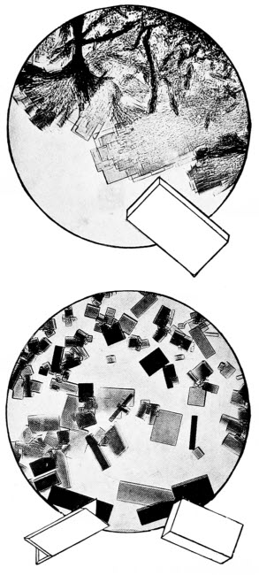
1. Crystals from Human Blood
It is often possible to identify the source of blood by means of crystals which can be obtained from it, for each race of animal has its own special form of blood crystal. The large crystal at the margin exhibits the typical shape.
2. Crystals from the Blood of the Baboon
These show some resemblance to human blood.
Now we may perform another operation upon ourselves, rather more painful than the last one but not very serious. We wish to examine some human blood, so we tie a handkerchief tightly round one of our fingers and with a clean needle—be sure that it is clean—make a puncture in the finger tip. The handkerchief bandage will prevent our feeling any pain. We must put the drop of blood we have obtained in the centre of a clean slide and examine it under the microscope. While the blood is still liquid, we shall see a number of circular discs floating about, they are very small being only 1/3200 inch in diameter. The centre of each disc appears darker than the rim, but this darker shade is only apparent. Using a term we introduced in our chapter on the lens, we may call the discs double concave. Now let us watch our objects for a moment and we shall notice that they begin to arrange themselves in chains, they appear like a number of draughtsmen placed one upon the other, our drop of blood is now beginning to clot. A simple experiment may be attempted at this stage; we must run a drop of water on to our little blood discs and watch carefully what happens. We shall see that they change their shape gradually, from being double concave they become flat sided, and this is not all, for they continue to change till both[170] sides bulge outwards and they may be described as double convex.
These little discs are known as red blood corpuscles. Their shape is some indication of the animal to which they belong; those of man, as we have seen, are circular and so are those of most of the higher animals, except the camel tribe, which has oval, red blood corpuscles. Birds, reptiles and fishes have corpuscles agreeing in shape with those of the camel. In size there is a great deal of difference between the corpuscles of various animals; a member of the deer family has the smallest and a creature related to our Newts, called Proteus, has the largest. The size of the blood corpuscles bears no relation to the size of the animal to which they belong, those of the frog measure 1/1108 inch, or nearly three times the size of the red blood corpuscles of man. Without much difficulty it should be possible to obtain other samples of blood and the red corpuscles should always be examined, needless to say the blood should always be in a fresh condition. In addition to the red corpuscles we may notice a few smaller circular bodies, they are the white corpuscles. If we have any difficulty in finding them in our own blood, we can examine another specimen of frog’s blood, in which they are more easily seen. It has been calculated that in a cubic inch of blood from a healthy human being there are eighty millions of red and a quarter of a million of white blood corpuscles. Although there is such an enormous difference in the sizes of red corpuscles[171] from various animals, the white corpuscles are remarkably constant, measuring about 1/3000 inch in warm-blooded animals and 1/2500 inch in reptiles. Sometimes, as we are examining the white blood corpuscles under the microscope, we shall notice that they behave in a similar manner to the Proteus Animalcule, which we described in our chapter on Pond Life. A kind of creeping motion takes place and the corpuscle loses its circular shape. In our own blood this movement lasts but a very few moments, in frogs’ blood, by keeping our slide moderately warm, we may witness the movement for some time.
It is perfectly easy to watch the circulation of blood in the foot of a frog. For this purpose we require a piece of apparatus known as a frog plate, this is merely a flat brass plate perforated with a number of holes, through which tape is passed to bind the frog down and having also a hole the size of the opening in the microscope stage, into which a circle of glass is usually fitted. Such a plate may be made of wood and will serve our purpose quite as well as the more expensive brass plate. We must bind our frog firmly, but not too tightly with wet rag, leaving one leg exposed. This free leg must now be fastened in such a manner that one of the webs between its toes comes over the opening in the plate, finally the toes must be carefully tied with string so that they remain apart and the web is fully expanded. Having fixed our frog in a satisfactory position let us examine his blood-vessels[172] under a fairly high power. We shall find that there are large and small vessels in his webbed foot; we will devote our observation to the small vessels, called capillaries, from the Latin Capillus, a hair, because of their small size. Probably the blood will not flow when we make our first examination and this points to one of two things, either the frog is bound too tightly or he has not recovered from the alarm he experienced at his treatment. In the latter event he will not be long recovering and his blood circulation will soon be in full working order; in the former case, we must loosen his bandages. In some of the very small blood-vessels we shall notice that the blood always flows in one direction, in others it does not appear to have any definite direction. In either case, however, we can see the red corpuscles flowing rapidly along the central stream of the blood-vessel whilst the white corpuscles travel much more slowly along the sides.
The medical man may be called upon to decide two questions concerning human blood; he may wish to know whether it is healthy and, in the case of certain crimes, he must be able to state positively whether certain stains are caused by human blood or not. Dealing with these questions in order, let us see how we would proceed. There are many people, far too many in towns who are described as bloodless, the expression is of course an exaggeration for no bloodless person could continue to exist. What really happens is that such people are deficient in red blood corpuscles. If we examine[173] two samples of blood, one from a perfectly healthy subject and one from a so-called bloodless subject we shall probably not be able to detect any difference between the two, but the experienced medical man will soon see that one sample has too few red blood corpuscles.
An unhealthy state of the blood is often indicated by the shape of the blood crystals. Let us see how we may obtain some of these crystals. If we add a drop of ether to our drop of blood upon the slide, and wait a few moments till the ether has evaporated, we shall notice when we examine our object again, under a high magnification, that a number of prismatic crystals have formed, especially towards the edges of the slide. Now in certain diseases of the blood, these crystals are no longer formed in their usual shape, thus pointing out to the medical man that something is amiss. Again there are many blood parasites just as there are external parasites of man. In the disease known as malaria, very small parasites are introduced into the blood by mosquitoes. Each of these little parasites enters a red blood corpuscle, divides up into many smaller individuals, causes the corpuscles to burst, then each little parasite attacks more corpuscles. Some of these little bodies are sucked up, along with the blood of malaria patients, by other mosquitoes and, if the insects are of the particular kind which spread malaria, the parasites complete their development within the mosquito. Patients suffering from that terrible African malady known as sleeping sickness,[174] have blood parasites resembling minute eels with long threadlike tails. These parasites are the cause of the malady and are introduced into the blood by flies, closely related to house flies. There are a very large number of blood parasites of one kind and another, so it is clear that a knowledge of blood is very important to the medical man.
From our remarks concerning the sizes of the red corpuscles in the blood of man and various animals, one might be excused from thinking that it would be quite easy for anyone with a little experience to recognise human blood. As a matter of fact it is very difficult, it is always a doubtful matter to rely upon size alone. We have mentioned blood crystals and these give us a slightly better clue to the origin of the blood, for the blood crystals of different animals vary in shape, far more than their corpuscles. Those of the guinea-pig, for example, are little four sided pyramids; those of the mouse, eight sided and so on. Some apes, however, have blood crystals very similar to those of man, so similar that the two may be confused. The microscopist who is compelled to give an opinion concerning the origin of a sample of blood, especially blood which is some days old, is faced with no light task.
Without the assistance of the microscope, medical men would never have discovered the cause of many of these insect-borne diseases, they could never have traced the development of the blood parasites in the bodies of insects. For many years[175] it was thought that the disease malaria was caused by the damp air of low-lying land, in fact the word malaria is derived from two Italian words meaning bad air. It was not till the advent of the microscope that the true cause of the disease was learned and the discovery was made that the only connection between the malady and dampness is that the mosquitoes which carry the disease can only thrive in damp situations.
Medical work with the microscope also entails a study of external and internal parasites of man other than those which infest the blood. Some of the larger human parasites we know only too well, they force their unpleasant attentions upon us from time to time, but fortunately they are not as a rule serious and are soon got rid of. The cleanest of us, in these days of universal travel, cannot avoid the visitations of the lively flea, he is at worst an annoying companion, but he has a cousin who is responsible for the passing of the germs of the dreaded plague from rats to man. Plague long remained a mystery, which, without the help of the microscope, would probably have remained unsolved to this day. Many of these apparently harmless blood-sucking insects may prove to be disease carriers, as medical knowledge and microscopic investigation is brought to bear upon them.
The majority of the smaller external parasites are beyond the reach of the amateur microscopist. A number of course are available to medical students,[176] but we write for the ordinary enthusiast and not for the specialist. Many interesting little mites, closely allied to the cheese-mites, cause skin diseases and the creatures themselves as well as their tunnels in the skin are interesting.
There is one little parasite which we nearly all of us carry without knowing it, and it makes quite an interesting object for the microscope. Its name is Demodex Folliculorum and it dwells in the sweat glands, especially those round about the nose. It is very minute and requires a high magnification for its examination. The adult is worm-like and tapering in the hinder two-thirds of its body, whilst the front third, to which is attached four pairs of short, fleshy legs, is stouter. From the eggs which may be heart or spindle shaped, little six-legged grubs arise; later these grubs change into the eight-legged adults. All the changes take place in the sweat glands, and the creatures live with their heads turned away from the gore of the gland.
To observe these strange little creatures it is only necessary to squeeze some of the white congealed perspiration from one of these glands on to a slide, coyer it with a drop of oil or xylol and spread out the object with a needle. By cutting down the light considerably by means of the diaphragm, we shall be able to distinguish all the stages of these little creatures which, unbidden, share our lives.
Other parasites come to us in our food and, of these, one of the most dangerous is known as Trichinella Spiralis and gives rise to a disease known[177] as trichinosis. We need not give a precise account of the life history of this interesting worm, it will suffice for our purpose to know that it finds its way into the bodies of human beings, in badly cooked pork or in improperly cured ham or bacon. The little worm is only about 1/25 inch long and, at first, dwells in the intestines of the pig. Each female produces upwards of fifteen thousand young and these pass into the blood of their host and thence to its muscles. Snugly coiled into a spiral between the muscle fibres, each young worm becomes surrounded with a lemon-shaped covering, which is exceedingly resistant. Suppose now a human being should happen to eat such pork, the juices of the stomach would dissolve the pork and also the little case enclosing the parasite. Once set free in the human body, the whole proceeding is repeated and, at last, the result is very severe muscular pains for the victim. The medical man is called upon to study such parasites as Trichinella, and they are not so uncommon as one might think.
In addition to animal parasites medical science demands a knowledge of many disease bringing fungi. One of the commonest is the fungus responsible for the unpleasant malady called ringworm. But we have written sufficient to show that the general practitioner needs his microscope always by his side. Not only so but very many instruments he uses in his work, though called by other names, are really microscopes adapted for special purposes.
Probably there are not many farmers who use a microscope and fewer still who use one to help them in their business, yet there are few people to whom one of these instruments would be more useful. Their seeds are often far from pure and the microscope will reveal the impurities which may consist of dirt and dust, or of other seeds, seeds which will grow into weeds and make the crop less valuable or, if present in large quantities, render it valueless. Agricultural plants become attacked by varied diseases which can only be studied under the microscope; insects also do their share of destruction and much may be learned about them when they are magnified. Fungi and insects not only attack crops but domestic animals as well. The microscope is an invaluable aid in studying the soil, in dairy work and in many other ways closely connected with agriculture.
That the testing of agricultural seeds is very important is shown by the fact that not very long[179] ago a deputation urged the Government to establish a National Seed Testing Station; no further plans have been made, however. Seed testing is very interesting work, every seed has its particular shape and markings and the student soon becomes absorbed in seeking for weed seeds among the collections he examines. A weed in the sense we use it here is not necessarily a harmful plant, it is a plant in the wrong place. For example a carrot growing in a field of turnips, though a useful plant would be a weed. When the farmer sowed turnip seed he did not do so with the object of raising carrots.
The only apparatus necessary for the study of most farm seeds is a powerful magnifying glass, one that will enlarge the seeds ten diameters or more. When beginning this work, a difficulty occurs at once for, without assistance from an expert, it is by no means easy to learn the names of the seeds one examines. The difficulty can be overcome to a certain extent if we know the names of flowers, for then we can collect the seeds from these flowers and we shall have properly named specimens as a guide. Beginning in this way, we shall soon find that the seeds can be arranged in groups and there will then be no difficulty in recognising say clover seed or grass seed, though much more experience will be necessary before we can say to which kind of clover or grass the seed belongs.
Many of these seeds are well worth studying,[180] whether we are interested in seed testing or not. The corn buttercup and the wild carrot have curious spined seeds; those of the larkspur when magnified appear to be studded with little shreds of paper. White and red campion, have kidney shaped seeds studded with warts and so similar to one another that the microscopist who can distinguish one from the other may consider himself something of an expert. Spurrey has lens shaped seeds with raised equator-like rims. Evening primrose seeds are curious because they are found in all sorts of shapes. The seeds of rib-wort plantain resemble miniature date seeds, others resemble minute bananas, some are perfectly round, others almost square; some have smooth shining surfaces which look as though they had been artificially polished, others again are wrinkled and deeply furrowed; but, most curious of all the common seeds are those of the cornflower, they resemble nothing so much as little shaving-brushes, with bright yellow bristles. Many profitable hours may be spent in studying the seeds of our common native plants, both wild and cultivated.
There are two specially obnoxious plants whose seeds may be mixed with agricultural seed, to the dismay of the farmer. We refer to Broomrape and Dodder, both of them unable to earn their own living and depending for their existence on the robbing of other plants. Broomrape usually attaches itself to the roots of Hazel, Poplar or Beech and steals its food therefrom, but its fleshy pink[181] stems and flowers may sometimes be seen in clover fields, then clover is its victim.
More common and more destructive is the little Dodder, a member of the Convolvulus family. Its seeds are very minute and when they germinate they give rise to a seedling not unlike a piece of wire. With one end fixed in the ground, the other waves about till it finds a clover plant round which it twines and not only so but it sends out suckers which microscopic examination shows, penetrate the stem of the clover to rob its food. By pulling a Dodder stem away from the clover we can clearly see a number of holes where the suckers have entered.
Fungus diseases and insects wage constant warfare on the farmer’s belongings. That we may better understand the structure of the disease-causing fungi we are about to examine, let us refresh our memories concerning that very common fungus, known as white mould and mentioned in an earlier chapter. The reason fungi cause damage to other plants, the one invariable reason, is that they, being unable to manufacture food for themselves, steal it from the plants on which they grow. Some of them are parasites and steal their food from living animals or plants; others live upon dead animal or vegetable matter and the white mould is one of the latter fungi.
In most of the fungi which concern us we shall find that there is a mass of minute, thread-like structures forming the main body of the fungus and[182] that, here and there, portions grow erect and bear spores. The spores, it is important to remember, serve the same purpose to the fungus as seeds to the flowering plants. They are blown or carried by insects or other agencies to suitable situations for growth, they germinate and form new fungi. They are smaller and lighter than the most minute and dust-like seeds, so that the slightest breeze scatters them far and wide. Let us compare the white mould with a mushroom; at first sight the two plants appear very dissimilar, in reality they are very similar to one another. The mould forms a thick felt of its threads over the substance on which it grows and mushroom spawn if carefully examined under a microscope will be found to consist of very similar threads, sealed together to form thicker root-like structures. The fungi, however, have no roots and these threads are strictly comparable with those of the mould. Here and there the mould sends a single thread into the air and each of these threads is terminated by a little ball which bursts eventually and sets free its contained spores. The same thing occurs with the mushroom; we have the upright growth, not of a single thread it is true but of a number, welded together to form a fleshy stalk; at the top there is, not a ball containing spores but an umbrella-shaped structure whose under surface is composed of a number of “gills” on which the spores grow at the ends of little stalks. If a piece of the mushroom stem be torn into its separate components and examined[183] under the microscope, its similarity to the more simple fungus is evident. One of the gills also may be carefully cut away and examined; the spores will be seen at the end of small forked stalks.
Having progressed thus far in our study of fungus structure, we may examine a few of those which cause damage in farm and garden. For the most part, the thread-like portion of the fungus grows within the plant attacked and only the spore bearing portions appear on the surface. There is one class of fungus, however, the Mildews in which practically all the structure grows on the surface, only a few small, unbranched suckers penetrate the plant attacked, for the purpose of obtaining nourishment.
Though of great interest to the microscopist, the potato disease is often the cause of serious loss to the farmer. Not only potatoes but also tomatoes are attacked. A potato plant suffering from the disease has irregular yellowish or brownish spots upon its leaves in the summertime. An examination of the lower surface of one of these spotted leaves will reveal a silvery white margin to each spot. This portion should be magnified with a fairly high power and care must be taken not to injure the diseased part of the leaf before it is examined. In cases of serious disease, from nearly every pore on the surface of the leaf fungus threads will be seen to issue. The threads are branched and, at the end of each branch, they have a special kind of spore. They look not unlike miniature leafless[184] trees and they give the typical silvery white appearance to the margins of the diseased spots. When the spores of potato disease germinate, the young fungus threads enter the leaf through a pore and for sometime afterwards there is no sign that the plant is diseased. On this account potato disease and many other fungus diseases are rendered more serious in that the farmer is not and cannot be aware that his crop is attacked till the disease has taken a firm hold. Eventually the potatoes themselves become brown, rotten and breeding grounds for bacteria.
A very common plant disease which makes a good study for the microscope, may be found in quantity upon shepherd’s purse, and as it also attacks cultivated plants of the same family, cabbages, cauliflowers and the like, it is of no little importance. In its early stages, the fungus looks like patches of thick white paint upon the plant and where the fungus grows the plant is invariably contorted. As the fungus matures, the skin of the diseased plant splits and a white powder issues. If some of this powder be highly magnified, it will be found to consist of chains of spores, six or seven in a chain. The spores break off singly and each one may start the disease in another plant.
The microscopist who hunts in garden and farm for fungus diseases, will assuredly meet with some examples of that large class known as “smuts.” They are so called on account of the black powder with which the attacked portions of the plants[185] become filled. The smuts are very important but are not of much interest as objects for the microscope, so we will pass them over for subjects of greater interest if of less importance.
Every farmer knows the familiar and destructive fungus known as “rust of wheat,” it is one of a large class of most interesting plants. The “rusts” are interesting to the microscopist on account of their structure and to the botanist because they cannot, like other fungi, complete their lives upon one plant. They derive their popular name from the fact that they look like patches of rust upon the plants on which they live. Some of the greatest living agricultural botanists have spent many years on producing races of wheat upon which rust fungus will not grow. Wonderful success has rewarded their efforts and conferred immense benefits upon farmers. In spite of this, however, we need not despair that we shall be unable to find a specimen for our microscope, though it is happily an undoubted fact that the disease is not so common now as a few years ago.
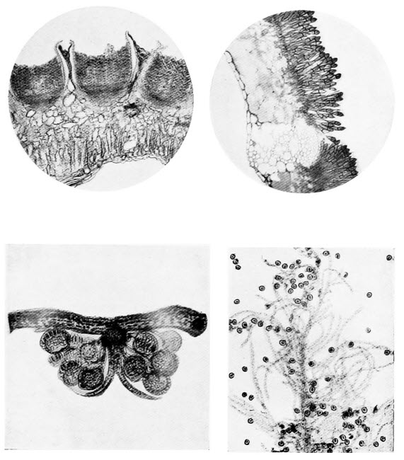
Photos by Flatters & Garnett
1. Cluster Cups
The spring stage of Rust of Wheat. Little orange cup-shaped growths on the under side of a Barberry leaf. They germinate on Wheat to form the summer stage of “Rust.”
2. Rust of Wheat
These little stalked spires are the winter stage of a serious disease of Wheat. In the spring they germinate on Barberry.
3. Pollen Grains on a Grass Flower
The feathery stigmas of grass flowers are beautifully adapted for catching and holding pollen grains.
4. The Lower Side of a Fern Frond
One of the brown outgrowths on the under side of a fern frond. The stalked spore cases are seen, protected by an umbrella shaped covering.
Rust of wheat fungus grows part of its time on barberry leaves and part on wheat. In the summer, if we examine one of the rust-like patches on stem or leaf of wheat we shall see that it consists of a dense bunch of small, short stalks each one of which is terminated by an oblong red-brown spore. If we keep another patch of the fungus under observation, we shall find as the season advances, that instead of the red-brown patch it has grown darker[186] and darker till it has become almost black. The microscope will show us that the structure of the spores has altered considerably. There is still the same bunch of stalks but they have lengthened somewhat and now each spore which terminates each stalk is divided into two parts by a wall across its narrow part. The walls surrounding the spores also appear thicker, as indeed they are. These are the winter spores, they fall to the ground eventually and there they remain, unharmed by frost or snow or rain, till the spring. In the spring they germinate and give rise, not immediately to another fungus, as might be expected, but to another kind of spore. Curiously enough these new spring-formed spores cannot grow upon wheat and unless they are carried by wind or some other agency to a barberry plant their existence is ended. Should they reach a barberry leaf, however, they will germinate, penetrate the leaf and grow for a period. Eventually the fungus appears on the lower surface of the leaf in beautiful structures called cluster cups. Under the microscope, one of these cluster cups forms a lovely object. The leaf skin is split and below the ruptured skin may be seen a flask-shaped hollow filled with chains of minute golden-yellow spores. The spores break away, one by one and favoured by fortune, are carried to a wheat plant where they germinate and give rise to the familiar rust. Any microscopist anxious for research has a life’s work before him in tracing the histories of this one class of fungi, should he feel inclined to shoulder the[187] burden. Very many cluster cups are known and very many rusts and all that is required is an enthusiastic mycologist, as the student of fungi is called, to put the pieces of the puzzle together, so to speak. It is not so very many years ago that the connection between the cluster cups of barberry and the rust of wheat was quite unthought of.
We cannot afford much more space to plant diseases, the farmer has other troubles and we must mention some of them. We cannot leave the subject, however, without a word concerning the mildews. As we have mentioned, they are curious because they dwell outside the plants they attack. Rose mildew is unfortunately all too common in every garden, it may be recognised as a white powder covering leaves and buds. Under the microscope, in the summer we shall find that it consists of a number of thread-like structures, not unlike those of the common white mould and that there are a number of erect chains of spores. Towards autumn, a further examination will show us many round dark-brown structures from which project a number of minute threads. These brown spheres are the winter stage of the fungus, designed to withstand inclement weather. In the spring, the spheres burst and set free a number of minute sacs, each one containing eight spores. The spores germinate on rose leaves and start the disease anew.
There will be no difficulty in finding mildews; they are all very similar to the rose mildew in general but they all differ in detail. The gooseberry[188] mildew for example, has a large number of threads running from its winter spheres and each thread is terminated by a little group of branches. The sacs which fall from the opened sphere in the spring, only contain four spores in this case.
The animal enemies of the farmer, so far as they concern the microscopist are more difficult to study. Many of them are internal parasites and to gain a real knowledge of their habits and life histories necessitates a good deal of rather unpleasant work for which the ordinary microscopist has neither the time nor the inclination.
In order to give our readers some idea of this work, let us take one of the commonest of all agricultural parasites and trace its life history whilst giving hints for its examination under the microscope. The common liver fluke is a worm which, in the adult stage, frequents the liver of some domestic animal, usually the sheep. A friendly butcher will probably be able to supply us with a specimen and, when we receive it, we shall probably dub it a very unwormlike creature. The worms form a large class in the animal kingdom and they do not all resemble the earthworm by any manner of means.
The liver fluke is a flat, almost leaf-like creature, it is not ringed like the earthworm and, under the microscope, we can plainly see all its internal organs. The fluke lays its eggs, each one enclosed in a little capsule, in the liver of the sheep. They are carried to the intestines and finally set free[189] along with the animal’s excrement. If then the eggs are blown, or carried by some means to water they will continue their development, on dry soil they cannot long survive. Each egg gives rise to a little organism which swims freely in the water; it is shaded like a blunt-ended cone when extended and is roughly oval when contracted. Its body is covered with little whip-like structures similar to those of the slipper animalculæ, and it is due to the lashing of these little whips that the creature moves through the water. If we found one of these young flukes in some pond water we might be forgiven for thinking it to be some near relative of the slipper animalcule. When our subject finds a living water snail it enters its breathing organs, becomes affixed to their walls and loses its covering of little whips. It becomes transformed into a shapeless mass which later develops into an elongated structure, quite unlike the free swimming creature which took shelter within the snail. Next, a migration is made to the liver of the snail where birth is given to, from fifteen to twenty, curious little heart-shaped organisms each with a tail about twice as long as its body. These little creatures escape from the snail and swim freely in the water for a time. Eventually they make their way to herbage growing by the waterside, affix themselves thereto and become surrounded with a hard coat capable of resisting the effects of hot sun or drying winds. Should this herbage be eaten by cattle, the apparently lifeless young fluke bestirs itself, loses its[190] tail and wends its way to the liver of its host, then the story begins again.
Having examined the adult liver fluke under the microscope, we shall probably wish to find both the free swimming young forms, and if we search carefully in ponds to which sheep have access we are likely to be rewarded. It is obvious that the life of a parasite such as the liver fluke is, of necessity, precarious. It is only chance or luck, or whatever one’s favourite term may be, that brings the egg to water, the young fluke to a snail, and the last free swimming form to herbage that will be eaten by a suitable animal. As usual in such cases, nature makes provision for emergencies by providing a large number of young, in order to insure that some at least may be able to complete their development. Owing to a series of changes, which we have omitted to describe for the sake of simplicity, each liver fluke egg may give rise to no less than three hundred and twenty of the final free swimming forms.
As we have remarked, the study of parasites is difficult but it is interesting. Very few of these creatures can complete their lives without living at the expense of two different animals. The liver fluke needs the water snail and some herb-feeding animal; there is another parasite which spends part of its life in the pig and another part in the grub of the cockchafer; a third parasite dwells for a time within the thrush, and for the rest of its time within the garden snail, and so on. Apart from the interest of the subject in itself, it brings us face to face with[191] the fact that many quite unrelated forms of animal life are essential to the well being of a number of parasites. To the farmer the subject is all important.
Insects of various kinds are all important in agriculture; most of them are harmful, some few are useful. They have, however, been dealt with in another chapter, so we will dismiss them here. The ticks are closely related, and anyone with access to a farm should be able to obtain some specimens. Whatever species we are able to obtain should be examined under the microscope. Their feet are always interesting, being furnished with powerful claws beautifully adapted to grasping the hairy coats of their hosts. Their mouth parts are quite unlike those of insects, and are always furnished with a number of backwardly directed teeth, which are useful for tearing flesh sufficiently to draw blood on which they feed.
Some of our readers will probably remark that entomology, or the natural history of insects, is really a branch of zoology and should be treated as such. We cannot pretend that they are wrong, but it is such a specialized branch that it merits separate treatment. Not many years ago insects, with few exceptions, were looked upon as harmless and often beautiful dwellers upon the earth. They afforded endless amusement to certain enthusiasts who collected them for their colouring or their odd forms. Recent developments of our scientific knowledge have shown us that the insect is, other human beings excepted, man’s most serious rival for the mastery of the world.
This state of affairs has been beautifully depicted by an American naturalist; his words described a by no means unlikely final scene on this earth of ours. He wrote: “When the moon shall have faded from the sky and the sun shall shine at noonday, a dull cherry red, and the seas shall be frozen over, and the ice-cap shall have crept downward[193] to the Equator from either pole, and no keel shall cut the waters, nor wheels turn in mills, when all cities shall have long been dead and crumbled into dust and all life shall be on the very last verge of extinction on this globe, then, on a bit of lichen, growing on the bald rocks beside the eternal snows of Panama, shall be seated a tiny insect, preening its antennæ in the glow of the worn-out sun, representing the sole survival of animal life on this our earth—a melancholy ‘bug.’”
There is probably no field more interesting for the microscopist than that provided by the insect world. Unlimited explorations may be made with the certainty of finding something new at every turn. Most people begin their studies of insect life with butterflies and moths; some folk to their loss never proceed further. We may well follow the usual course, and make a butterfly our first study.
Any butterfly or moth will do for our purpose, any one with coloured wings, for some have clear wings like those of the bees and wasps, but they are not very common, so that we shall probably find a suitable specimen at the first attempt. The more highly coloured the specimen, the more attractive it will appear under the microscope. After killing the insect, and not before, we may proceed to study it. Killing may best be accomplished by means of a killing bottle, failing this a hard nip on the body, between thumb and finger, will do, but it must be no half-hearted proceeding or the insect will be injured without being killed.
Having removed a wing and placed it on the stage of our microscope, we must examine it by reflected light, for it is not transparent. This may be accomplished, if we are using artificial light, by raising the source of illumination well above the object, so that the light strikes it at an angle of about forty-five degrees; by daylight reflected light is easily managed. If we have never previously examined a similar object we will be surprised at its appearance. All the beautiful reds and blues, yellows and greens which comprise the brilliant livery of these insects are seen, under the microscope, as hundreds of minute scales which overlap one another like tiles on a roof. A higher magnification will show that each scale is roughly flask-shaped, and that its narrow end fits into a little socket in the wing proper. When the scales are rubbed from the wing, nothing remains but a transparent substance traversed by veins—to the microscopist the scaleless wing is of little interest; to the entomologist it is important, for the moths, at anyrate, are arranged into families largely according to the arrangement of the veins of their wings.
Many other wings may be examined with advantage; gnats, for example, are clothed with scales of varied shape, some hair-like, some forked, some resembling a sickle, and some disc-shaped; these forms, by the way, do not all occur upon the wings, but are found upon the head and other parts of the body as well. The wonderful gauzy, iridescent wings of dragon flies are interesting; those of[195] various flies worth examination also those of bees on account of the clever device for uniting the front and hind wings during flight. On the front edge of the hind wings the microscope will show us a row of minute hooks. When the bee makes a flight, it hooks its hind wings to a ridge on the hinder edge of the fore wings, so that for flying purposes it has, to all intents, two wings instead of four.
Having examined all the wings we can find for the time being, we may turn our attention to mouths. The mouth parts of insects are not only interesting but important; one of the first things an economic entomologist does with a new specimen is to examine its mouth parts. The mouth will tell us what manner of feeder its owner may be. Some insects have sucking mouths, and they must feed perforce on liquids; others have biting mouths, and they are likely to do damage to crops by eating them. Then there are lancet-like mouths and mouths which are a compromise between biting and sucking ones. The subject, however, is somewhat complicated, and entails a knowledge of insect anatomy, so we will merely deal with a few easily understood examples. Our butterfly has a sucking mouth; it is known as a proboscis, and may be found, coiled like a watch spring, beneath its head. There is no trace of anything in this mouth capable of biting or even piercing the most delicate structure. The house-fly is also possessed of a proboscis though of different design. Though a dangerous, disease-carrying insect, it can do no harm with its[196] mouth. The partiality of the house-fly for sugar is well known, and it is interesting to learn how, with its soft fleshy mouth, it can satisfy its cravings. Let us watch one at work on a lump of sugar through our pocket lens. If we look carefully we shall notice that the fly, as he thrusts his proboscis here and there, emits from it a little drop of liquid; after a momentary pause the liquid is sucked up again, it has dissolved a little of the sugar, and the fly enjoys the sugar-laden liquid.
People frequently state that they have been bitten by a house-fly—a sheer impossibility. What really happens is that they mistake the very similar stable fly for the house-fly. If one of these insects be captured and examined, we shall find not the soft fleshy proboscis of the house-fly, but a cruel looking, awl-like mouth easily able to penetrate the human skin. Certain tropical flies, known as Pangonias, have such formidable and lengthy piercing mouths that they can penetrate thick clothing and puncture the skin below.
Microscopists who care to follow up the study of insects’ mouths will know that they are accumulating really useful knowledge. Those who do not desire to go so deeply into the matter may well spare a few moments for the examination of the green fly mouth, a needle-like piercing organ which is thrust into plants for the purpose of sucking their sap. The mouth of the gnat is a more difficult subject for the microscopist, though no less interesting; it may be compared with the same organ of[197] the green fly, for it is used somewhat similarly, with the difference that it sucks blood and not plant juices. It may be well to mention here that the females alone suck blood, but it is easy to distinguish the sexes, for the antennæ or feelers of the females are thread-like whilst those of the males are feathered. A few adult insects have no mouths, for they never feed during their short lives.
Caterpillars of all kinds and also beetles, grasshoppers, cockroaches and the like have biting-mouth parts, and the student, who is not well versed in insect anatomy, will learn more by watching one of these insects partaking of a meal than by trying to discover the uses of the various parts with the aid of a microscope. Caterpillars as a rule are not shy feeders, and a pocket lens will show their sickle-like jaws in full play. The grubs of house-flies are worth examining; they are soft and fleshy except for a pair of horny hooks which are used to tear up the food material. There are, however, so many different mouths we cannot describe even the typical ones, but the microscopist will soon discover those of special interest.
The feet of insects do not show so much variation as their mouths, nevertheless they will afford ample material for many hours of study. Our butterfly, which is now but a remnant, will provide our first object. The design of its feet will depend, to some extent, on the species of insect, but they will certainly be clawed. Other insects with clawed feet—beetles, bees, and wasps—may be examined, and[198] we shall see that there are minor differences amongst them though their general plan is similar. Sometimes we find a simple pair of claws on each foot, in other cases each claw has a little spur, whilst spiders, which, by the way, are not insects, have comb-like claws. The foot of the house-fly is not only provided with a pair of claws, but also with a soft fleshy pad, by means of which it is enabled to climb window panes and similar smooth surfaces. If we are fortunate enough to obtain a specimen of a louse, human or otherwise, we must not fail to notice its strong grasping claw, used for taking a firm hold of the hair of the creature on which it lives. Such objects are better examined under a high magnification, along with a hair, then the actual method of grasping may be observed.
The feelers of some insects are interesting; those of gnats we have already mentioned, but they may be examined in detail. Those of beetles are of very diverse form; some are thread-like, some clubbed, some fan-shaped. Moths, too, have many curious designs to show. Some of these feelers, when highly magnified, may be seen to be pitted—hundreds of little sunken areas are scattered over their surfaces, and it is probable that they are connected with a sense of smell. In that case the feeler is a more important organ than one might surmise from its popular name.
The hairy clothing of insects need not delay us long. Most interesting of all are the feathered hairs[199] of certain bees. In our chapter on botanical work with the microscope we mentioned the feathery stigmas of grass flowers and we also stated that they took that form, so that pollen grains blown to them would be entangled in their branches. The hairs of many bees are feathered for a similar reason, they gather pollen and the pollen adheres to these “feathers” much more readily and in much greater quantity, than it would to simple, unbranched hairs. Some bees collect no pollen and, from them, feathered hairs are wanting.
Any microscopist who has followed us thus far, will have a fair idea of the structure of a number of insects. In every case, where possible, comparisons should be made between similar organs of different insects and the investigations may be made more interesting by observing the habits of the insects and trying to discover reasons for the differences in structure. It is safe to surmise that there is a reason in every case. There are many other interesting subjects which we have not mentioned, the legs of insects—running legs of ground beetles, digging legs of mole crickets, swimming legs of water beetles and the wonderful pollen-carrying legs of bees. Then again, the eggs of many insects are of surpassing beauty in shape, they may be round, oval, oblong, nearly square and almost needle shaped; some are smooth and shining like burnished metal, others beautifully sculptured; some resemble miniature birds’ eggs, others are not unlike the seeds of plants.
Many insects are too small to be cut up into[200] their various parts, legs, feet, wings, etc., by unskilled hands and they must be examined whole. Perhaps you may think that an insect will be too big an object for your microscope. Indeed there are some insects which measure nearly a foot in length, but there are, on the other hand, beetles no longer than one hundredth part of an inch. When we examined the house-fly it is not unlikely that we found some minute creatures living upon it. None of these is likely to be an insect, but as they are closely related we may mention them here. Beneath the wings of the house fly there are often minute, red, six legged young mites, all eagerly sucking the juices of their host. Because they are six legged we may be led to think that they are insects, for the entomologist knows that the true insect, in its adult form, never has more than six legs. These mites, however, later in life, drop from the fly and by developing another pair of legs appear in their true colours. Various other mites, including cheesemites, may be found clinging to the house-fly, in fact it is by the aid of these insects that cheesemites are often carried from cheese to cheese. One of the most curious parasites of the house-fly must be sought upon its legs. If the search is successful, curious, reddish-brown creatures, armed with formidable pincers, and strangely reminiscent of miniature lobsters, will be found clinging thereto. They are called chelifers and their home is the manure heap, so that their presence shows us only too well where our friend the fly has recently disported himself. His next visit was probably to our food.
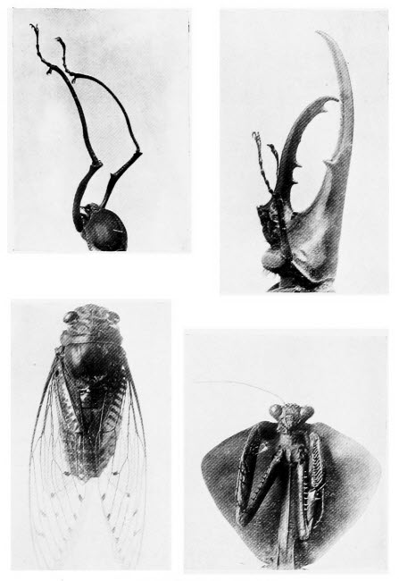
1. The Head of a Beetle
A remarkable beetle, with enormously developed fore-legs. The object of these long legs is not known.
2. The Head of Hercules Beetle
This beetle, whose head resembles a lobster’s claw, is said to carry his wife from place to place.
3. A Cicada
One of the noted singing insects. Kept as a pet in some countries, voted a nuisance in others, the cicada is the medium of much romance.
4. The Head of Mantis
This illustration clearly shows the cruel rat-trap-like front legs of the rapacious mantis.
If we can find a swiftly running stream within easy reach a little time may be spent in searching the submerged rocks and plants for the miniature stages of the buffalo gnat. This insect, which is known to scientists as Simulium, has a most interesting life history. Its popular name is derived from its hump-backed appearance and its supposed resemblance to a buffalo. The female lays her eggs on a rock or reed, just covered by running water, she never lays them in still water. The greenish-brown, club-shaped grubs which come from the eggs are curious and they will repay examination under a low magnification. At the more blunt end of the creature there is a large sucker; it uses this as a foot to support itself in an upright position. If we examine our specimen under water we shall see that its horny head is decorated with a pair of fans, each one composed of about fifty threads. These fans open and close with a rhythmic movement and, in doing so, attract small floating water plants to the mouth. Just below the head there is a single leg with a sucker foot. When the grub walks, it does so by a looping movement, holding fast to its support with fore and hind suckers alternately.
The next stage in the life of the young buffalo gnat is even more curious. On the surface of some submerged leaf we shall probably be able to find a number of the slipper-shaped pockets made of closely woven silk, in which the insect spends the[202] final portion of its life before turning into the perfect fly. Within some of the pockets we shall find the creature itself and it must be studied. Its head is still ornamented with a pair of fans, but in this case they are gills by means of which the insect breathes and not food scoops, for it has reached a stage when food is not taken. On its tail end there is a hook, by means of which it anchors itself to its slipper-shaped pocket. Probably we shall find bubbles of air collecting round some of the insects, within their pockets; as the time approaches nearer and nearer for the change to the perfect insect to take place the bubbles grow larger and larger. Eventually the fly emerges within a bubble, shakes it free from the pocket and floats to the surface of the water without wetting its wings.
The pond will supply insects quite as interesting as the running stream. Here we may find the eggs of Caddis flies, enclosed in jelly-like envelopes, rope-like, horse-shoe shaped or simple masses. The caterpillar cases of these insects should be slightly magnified and examined, for they are marvellously constructed of shells or sand, pieces of stick, leaves or other vegetation.
We may come across the young stages of the common gnat. Its eggs are very small and when the mother gnat lays them on the surface of ponds she glues them together so that they form what is known as an “egg raft.” The “raft” we shall notice, if we examine it carefully, is composed of a large number of eggs; each egg is elongated,[203] pointed at one end and blunt at the other. Every egg is arranged with its blunt end downwards for, from that end, the larvæ gnats make their way into the water. A larva, it may be explained, is the stage in insect development which follows the egg; the next stage is known as the pupa. When the eggs are first laid they are white but, before they hatch, they become darker and darker, till they are almost black. The larvæ which hatch from the eggs we must examine under our microscope and also in the water—they may easily be kept in a small jar of pond water. If we study their habits carefully, we shall observe that they float almost at right angles to the surface of the water and that, while doing so, the tip of a little peg-like outgrowth on the tail end of their bodies is thrust out of the water. The little peg is the breathing organ of the gnat larva and the flaps which open and close at its tip are worth examination. In a few days, the denizens of our pond water will change their appearance and become comma shaped. On their heads we shall see a pair of curious horns, which project out of the water as the creatures, which have now become pupæ, float at the surface. The horns are breathing organs. As we examine these pupæ, day by day, we shall see the various parts of the complete gnat as they develop within the body of the comma. Finally, under the microscope, we can trace all the parts of the gnat. Darker and darker the little creatures become, as development proceeds; at the same time they become less active and less comma[204] shaped. At length the time comes when they straighten their tails somewhat violently, their skin splits along the back and out comes the perfect gnat. We can use him for further microscopic work, so we will not let him go. If he be a male, his beautiful feathered feelers or, if a female, her thread-like feelers will make good objects for us, the scales from head and body, the wings and feet are all worth the time we may spend in examining them. The mouth parts, too, are interesting but rather complicated.
Many insects are capable of emitting more or less musical sounds. In some countries these so-called singing insects are kept as pets, in other countries the same insects are voted a nuisance; it is all a matter of taste. Of all musical insects, the most noted is the Cicada and its sound organs are easily seen; they occur only in the male, for the female never sings. The Cicada belongs to the same great order of insects as the green fly, which we have already mentioned. There is one British species, and our readers who visit the New Forest may come upon it. On the under side of one of these insects the beak, very similar to the beak of the green fly, may be plainly seen. On either side of the insect, just below the bases of the wings there are two nearly round discs. These discs cover the sound organs, which are two ear shaped membranes. By means of muscles the insect can cause the membranes to vibrate and thus produce the sound which once heard, can never be forgotten.
More easily found insects, in this country, at any rate, are the cricket and the grasshopper. The cricket we all know is a persistent songster. Let us examine him closely. We shall find, that the house cricket has two pairs of wings; the fore wings are leathery, the hind wings membranous. If we watch a male in the act of chirping, the male crickets like the male Cicadas are the songsters, we shall observe that he moves his wings slightly. If now we examine a dead male, we shall find, on the under surface of the forewing, a rough patch. Let us examine this patch under the microscope, it reminds us of nothing so much as a file: it is a file in fact, and sound is produced by rubbing this rough file against a ridge, which we can easily see, on the upper surface of the hind wing.
It would be a useless accomplishment for the cricket to be able to sing, if there were no ears to hear its song. Nature has arranged that his song shall not go unheard and if we examine a female cricket, that is to say a cricket which has no sound-producing apparatus, we shall find an oval depression, covered by a membrane, on each of her front legs; these are her ears and they enable her to hear her mate calling to her.
We have often heard the song of the grasshopper as we have walked through the fields and he too will occupy our time for a few moments. When he sings, he kicks his legs rather violently and this gives us a clue to the situation of his vocal organs. The inside of each of his hind thighs is ridged and[206] the edge of each ridge is, as we can see if we magnify it, rough like a file. This file-like ridge is rubbed against a smooth ridge on the edge of the fore wing and the result is the familiar note of the grasshopper.
This insect also has ears, but they are not easy to find unless they are pointed out to us. If we examine a grasshopper we shall see that its body is divided into three parts: a head, a solid portion from which the wings and legs arise, the thorax and a portion (the abdomen) made up of a number of rings or segments. On the sides of the first of these rings, counting from the forward end, we shall find small depressions covered with membrane, these are the ears. It is curious that although many of the grasshoppers cannot give out a note, so far as human ears can detect, they nearly all have ears; maybe there is a grasshopper song which only grasshoppers can hear.
Very many other insects have sound organs, but they are nearly all constructed on the same plan. It may seem surprising that sounds can be produced by these simple means. Sound is really caused by waves in the air and these insect vocal organs set up rapid sound waves, by their vibration.
The microscopist should never be at a loss for objects derived from the insect world: it is impossible to walk without treading upon some six legged wayfarer. The wing cases of beetles are often of rare beauty, some on account of their sculpturing, some because of a mantle of scales.
In our greenhouse and garden we can find mealy[207] bugs, curious little powdered insects which do an enormous amount of damage. The fringe wings, or Thrips, are common and destructive; examine their curious feet and their beautifully fringed wings, the sight will repay you. And lastly, if you number an insect enthusiast amongst your friends, enlist his aid in gathering objects for your microscope.
It is always surprising that the majority of microscopists never dream of examining any of the hundreds of beautiful objects which can be found by the seaside, in the course of an afternoon’s ramble. That every pond will contain ample material for study, the microscopist knows instinctively; insect life and plant life also he studies, but the microscope is generally left at home when a visit is paid to the seaside. A rocky coast is better than a sandy one, for rock pools yield many objects, and the warmer southern waters of our coasts are better than the colder northern seas, but the microscopist who finds himself on a northern sandy coast need not despair, if he search diligently he will find material enough to occupy him for many a day.
Nearly every rock pool will provide one or more Sea Anemones; it is hardly necessary to describe these “flowers of the sea” as they have been called, they are such familiar objects and the brilliant colouring of some of them makes them highly attractive.[209] In many respects Sea Anemones resemble the Hydra, one of the pond dwellers, they are rather more highly developed, however. Any Sea Anemone will serve our purpose because we are about to examine the little darts with which its tentacles, and even its body, are armed. If we find several different kinds of Anemone, we must take the most transparent we can find and also a small specimen; we can examine the larger, more opaque ones later. Having transferred our specimen to a small jar, containing but a small quantity of sea water, we wait till it has recovered from its transfer and spread its tentacles, then it must be killed by one of the methods suggested in our concluding chapter (see p. 306). One of the tentacles must then be snipped off with scissors—some people cut off the tentacle without killing the Anemone and the animal does not appear to suffer a great amount of inconvenience, in fact a new tentacle soon grows to take the place of the old one. We do not recommend promiscuous vivisection. The tentacle is placed on a clean slide, a cover slip placed over it and pressure is applied. An enormous number of little thread-like darts are pressed from all parts of the tentacle. In some cases, little oval capsules are squeezed out and, in the capsules, the darts may be plainly seen, coiled up. On applying pressure to a capsule, the contained dart will shoot forth, much as does a glove finger turned inside out, when we blow violently into the glove. These little darts are of the greatest interest to the microscopist; they[210] vary in shape according to the kind of Anemone, as we shall find if we try this experiment with various Anemones. Some of them are straight with stiff bristles at their bases; some have backwardly directed barbs at their tips; others are apparently jointed, forming a zigzag, with a short length of the dart going from left to right, the next short length from right to left, and so on to the tip. It is marvellous how the darts can be accommodated within the capsule, for the average length of the latter is but 1/300 inch whereas its dart may measure 1/8 inch. These little threads contain a poison capable of paralyzing any moderate-sized fishes which they touch.
Have you ever seen a “comb bearer” or as it is often called, a “marble bleb?” Probably you have though you may not know its name. Sometimes it occurs in rock pools, though more often it is found in one’s shrimping net and occasionally it is washed up by the tide, but it does not live long out of water. The “marble bleb,” as its name denotes, is an almost globular mass of soft, transparent jelly. It is practically colourless, with the exception of eight bright coloured bands which run from end to end of the animal. To the naked eye, this little denizen of the sea is of rare beauty: as an object for low-power microscopy it is entrancing.
When magnified, the bright bands are seen to be composed of rows of flattened outgrowths. If our specimen is small enough to be examined in water,[211] its real beauty can only be seen in this manner, we shall observe that the flattened outgrowths act like paddles, sometimes they work all together, sometimes independently of one another and this fact explains the marvellous evolutions of the “marble bleb” in water. Now it shoots forward in a straight line with some rapidity, now it rolls over and over and swims onward while doing so.
In the sun, it displays glorious iridescent colouring. At the hinder end of the “bleb” we notice a pair of hollows: from these, as we watch it swim, we shall see it suddenly shoot out a pair of long feathery tendrils and they may be withdrawn into the hollows as suddenly. We must make a point of examining the “marble bleb,” it is one of the gems of our coasts.
Superficially the common, sponge-like “Dead man’s toes” or, to give it its more pleasant title, “Mermaid’s fingers,” is a very drab affair. It is a dirty-brown lobed, spongy mass with a leathery skin; when removed from the water it loses all semblance of shape. In sea water, however, if we examine it carefully, we shall see that it is studded with beautiful little flower-like creatures, each one resembling a miniature Sea Anemone. Examined, in water, under a low power of our microscope we can see the water current flowing through the channels with which it is perforated, after the manner of a sponge. If, now, we take a dead specimen and cut it up, placing a small portion on a slide and shredding it with a pair of needles, we shall find, when[212] we magnify the result of our work, that there are a number of minute mineral spikes, called spicules, and very beautiful objects they make for the microscope. There are many sea-side creatures, which we may find, closely related to “Mermaid’s fingers,” they may all be treated in the same way for they will all yield spicules which will repay us for our trouble. All these spongy organisms are not provided with spicules as ornaments, though one might be forgiven for thinking so, seeing how decorative many of them are. Their presence is necessary to strengthen the spongy material.
The specimen we have just examined is not one animal but a colony of very minute animals. These colonies are very common, not only in salt but also in fresh water. They serve a useful purpose, for the creatures composing them are so minute that they would fare badly did they dwell alone. Dwelling together as they do and each one contributing its share to the building of the home they appear to thrive to a wonderful degree. The coral islands are built, as to their foundations at anyrate, by millions of very minute animals, living together in colonies.
We mentioned the sponges a moment ago and many of them may be found around our coasts; not the household sponges we know so well, but much smaller, though equally interesting, colonies. Like the better known sponges, those which we find on our shores are perforated with a number of holes[213] through which the water is driven by means of little whip-like structures which line the cavities. Professor Grant has graphically described his impressions at witnessing this water current for the first time. “I put a small branch of the Spongia Coalita,” he writes, “with some sea water in a watch glass, under the microscope, and, on reflecting the light of a candle up through the fluid, I soon perceived that there was some internal motion in the opaque particles floating through the water. On moving the watch glass, so as to bring one of the apertures on the side of the sponge fully into view, I beheld, for the first time, the splendid spectacle of this living fountain vomiting forth from a circular cavity an impetuous torrent of liquid matter, and hurling along, in rapid succession, opaque masses, which it strewed everywhere around. The beauty and novelty of such a scene in the Animal Kingdom long arrested my attention; but after twenty-five minutes of constant observation, I was obliged to withdraw my eye from fatigue, without having seen the torrent for one instant change its direction, or diminish, in the slightest degree, the rapidity of its course. I continued to watch the same orifice, at short intervals, for five hours, sometimes observing it for a quarter of an hour at a time, but still the stream rolled on with a constant and even velocity. About the end of this time, however, I observed the current to become perceptibly languid ... and in one hour more the current had entirely ceased.”
Frequently the sponge we examine may be found to be studded with many yellowish spots; closer examination will show that these spots are composed of very small jelly covered eggs. Later these eggs find their way into the cavities of the sponge and are forced therefrom in the currents of water. Each of the young sponges thus expelled is furnished with a covering of little whips, by means of which it swims about till it can find a suitable spot on which to anchor and complete its growth.
The Sea Anemone, which has already provided us with objects for our microscope, has many near relatives which we must make a point of examining, while we have the opportunity. Many of these creatures, or rather their colonies, for they do not live singly, are to the naked eye, strangely like seaweeds. A number of them are moss-like and may be found on wooden breakwaters and similar situations when the tide is low; they should be collected and examined and, to see them at their best they should be examined under water. It is hardly necessary to describe any one of these colonies in detail, for they are so numerous that the one we described might not come into the hands of our readers for a long time. In general characters they are all somewhat similar so we will confine ourselves to generalities. For the most part, the stems and branches of these colonies are of the thickness of thread. As we watch them under the microscope we shall see that they are studded with little cups and, presently, from each little cup there appears[215] a little tuft of tentacles which is waved about in the water. Each member of the colony is similar to its neighbour and each one, again, is very like the fresh water Hydra with which we are familiar.
Of all the common objects of the sea shore one of the commonest everywhere is the sea-mat. Nine people out of ten or, we might safely say that everyone who had not learned its true nature, would guess it to be a seaweed. As we find it washed up on the beach it is almost the colour of sand, somewhat rough to the touch, whilst its whole surface is pitted with minute holes. The sea-mat, when dry as we usually find it, is a remnant of a colony of sea dwellers very similar to those we have just described. From each little hole, in a living specimen, which we can find without much difficulty, there appear the familiar tentacles; each hole is the home of a minute hydra-like animal.
Hooke, whom we mentioned in our chapter on the History of the Microscope, though a careful observer, was quite misled by the sea-mat; he thought it was a seaweed, for he wrote: “I have not, among all plants and vegetables I have yet observed, seen any one comparable to this seaweed. It is a plant which grows upon the rocks under water and increases and spreads itself into a great tuft, which is not only handsomely branched into several leaves, but the whole surface is covered over with a most curious knot of carved work, which consists of a texture much resembling a[216] honeycomb, for the whole surface on both sides is covered over with a multitude of very small holes, being no larger than so many holes made with a pin, and ranged in the neatest and most delicate order imaginable, they being placed in the manner of a quincunx, or very much like the rows of eyes of a fly, the rows or orders being very regular which way soever they are observed. These little holes, which to the eye look round, when magnified, appear very regularly shaped holes, representing almost the shape of a round-toed shoe, the hinder part of each being, as it were, turned in, or covered by the toe of the next below it. These holes seemed walled about with very thin and transparent substance, looking of a pale straw colour, from the edge of which, against the middle of each hole, were sprouted out four small, transparent, straw-coloured thorns, which seemed to protect and cover those cavities.”
As a well-known author has remarked: “This is really a wonderfully faithful description of the common sea-mat, and one cannot help picturing the surprise and delight of old author Hooke, could he have seen a portion of a living colony under a modern microscope.”
One of our finds may be the “Bird’s head.” It is a branched form, quite unlike the sea-mat but it is of even greater interest. Under the microscope, we shall see the many waving tentacles, but another feature is sure to attract our attention, a feature which is responsible for the popular name of the[217] colony. On the outside of each cavity containing a member of the colony there is a structure which resembles nothing so much as a hawk-like bird’s head atop of a long neck. While the tentacles wave in the water, the bird’s head snaps vigorously, moved here and moved there. The birds’ heads, which might be mistaken for parasites stealing food from the waving tentacles, really perform the useful function of keeping them clean and warding off creatures which might do them harm.
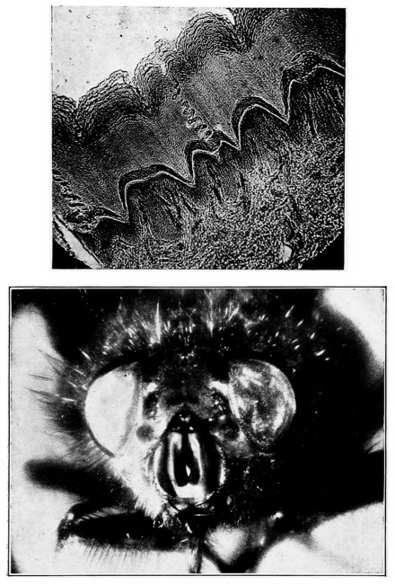
By the courtesy of Messrs. F. Davidson & Co.
1. A Section of Human Skin
The corkscrew-like pores, leading from the sweat glands to the surface, are plainly seen in the section.
2. The Face of a Fly
A wonderful photograph, at 15 inches, taken by the micro-telescope. Notice the very large size of the eyes relative to the rest of the head.
Now let us pass to quite different though equally common sea shore animals, the star fish. There are very many kinds but the common star fish will serve our purpose well. We may make a beginning by examining his back under a low magnification and observing that it is protected by a number of hard plates which form a very efficient armour. At the point where two of the rays (the finger like structures) arise we shall notice a small flat plate, this too is worth a moment’s inspection, for it is the water pore through which the star fish takes in water.
The under surface of the star fish shows us of course its mouth in the centre of its body, the soft fleshy suckers which cover the rays, with the exception of a narrow line down the centre of each one. At the tip of each ray there is an eye; it may easily be distinguished by its bright red colour and microscopic examination will show us that it is quite unlike any of the other eyes we have examined. Over each eye there is a little tentacle; these little[218] tentacles may be called the noses of the star fish, for, by means of them it is able to smell. They are as unlike our idea of a nose as are the little pits on the feeler of the cockchafer which we examined, yet these noses and ours all perform the same duties. Our time will be well spent if we devote some of it to a search for others less common star fish. Some of them are really beautiful and whatever specimens we come across can be compared with the common variety which is everywhere.
Closely related to the star fish are the sea-urchins. The relationship may not be apparent at first sight but a careful study of an urchin and a star will reveal many points of similarity. Our object in these pages, however, is to find material for our microscope and not to unfold the relationships of various members of the Animal Kingdom. When we have learned to cut sections, we may try our hand at the spine of a sea-urchin, it is an object well worthy of study. The hard shell of the urchin may be examined under a low magnification, we shall see then that his armour is far more highly developed than that of the star fish.
Another near relative of the two animals we have just described is the sea slug or sea cucumber. Though an article of commerce of some importance in the far East, the sea slug is not so common on our coasts as its relations. We must make a point of finding a specimen, however, for it provides us with one of the most remarkable objects for our microscope that could possibly be imagined. One[219] species, which goes by the name of Synapta Inhærens, is the one most worthy of examination. We must describe the creature first of all so that we may know what to look for. It is aptly named sea cucumber for it is not unlike that fruit—yes! fruit is correct, though the cucumber is more often called a vegetable. The animal’s skin is tough and leathery and at the head end there is a fringe of feathery tentacles. The sea cucumber must be looked for amongst sea weeds or, maybe, he lies buried in the sand, with only his fringe of tentacles on view. A friendly fisherman will probably aid us in our search.
Having found our creature we must examine his leathery skin under the microscope. To the touch it is evident that it is studded with some flinty matter, but the microscope alone can show us the amazing beauty of this armour. Under a low magnification, we can see, dotted over the leathery skin, some nearly circular plates to each of which is attached a little anchor. Now, from a dead animal of course, we must scrape away some of these objects and examine them with a higher magnification. Even the hardened microscopist will be delighted when he sees the armour of the sea cucumber for the first time. Each anchor is hinged to a little plate, each little plate, nearly circular in outline, is perforated with seven holes, six round the circumference and one in the centre and every perforation has a toothed margin. So perfect are these minute plates and anchors, that the most[220] intricate man-made machinery could not have turned them out more perfectly to pattern. They are precisely similar to one another in size and shape; as objects for the microscope, even the sea with all its store of wonders, can offer us nothing more marvellous.
We may number the sea mouse amongst our treasures of the sea-side. Though called a mouse, on account of its curious movements and partly perhaps because of its appearance, it is really a worm. It does not appear to have the slightest resemblance to the common earth worm, nor to the liver fluke which is described on another page, nevertheless, it is related to both these creatures. The raiment of the sea mouse is gorgeous in the extreme; on its back is soft brownish hair, its sides are clothed with yellow and green hair, displaying a wonderful iridescence and amongst the hair on the sides there are many stiff brown bristles. Of the covering of the sea mouse it has been said: “It is as if all the hues of the rainbow were collected there, making this remarkable animal a living jewel, and truly worthy of the name of Aphrodite, the Queen of Beauty.” The bristles of this creature we must examine microscopically, they vary in structure according to the kind of sea mouse, for there are several kinds, but in some of them they are formidably barbed, in all of them they act as a protection.
Many other worms, a number of them un-wormlike in appearance will claim our attention but we[221] cannot devote more space to them here. There is the common shell-binder, a curious worm, which builds for itself a still more curious shelter of broken shells. Another worm, Serpula by name, also lives in tube-like structures of its own manufacture and is remarkable in that a row of nearly two thousand seven-toothed hooks run along its back and all these thirteen thousand odd teeth are there merely for the purpose of holding Serpula in its tubular home.
Much interesting work may be done at the seaside in studying the young stages of various familiar creatures. This work, however, necessitates the keeping of the adults in an aquarium; for the young ones, in most cases, are so unlike their parents that, if found swimming about on their own account, they would never be recognised. Young barnacles, for instance, have six legs, a tubular mouth and a single eye. At a later stage they might be mistaken for young shell fish; they possess a shell, not unlike that of the mussel, containing, however, not a soft, fleshy mollusc but a six legged creature which swims through the water in jerks, after the manner of a water flea. Strangely enough, it is now provided with two stalked eyes like those of a crab, whilst of mouth parts it has none, or they are so imperfectly developed as to be useless for feeding. Soon this active youngster settles down for the rest of its life and becomes a sedate and sedentary barnacle, with one imperfectly developed eye and a mouth capable of feeding to good purpose.
Young crabs are equally curious and also totally unlike their parents, but these curious creatures are hardly accessible to those who only pay a flying visit to the sea-side. As we have remarked, an aquarium is a necessity and to keep marine animals inland is a feat beyond the powers of the ordinary mortal.
The Sea Lemon or Doris is a curious little creature, worthy of examination. Its habit is to feed upon sponges and strange diet it is, for we remember that all sponges are fortified with hard flinty structures, called spicules. This habit of the Sea Lemon is of use to the microscopist, for the stomach of the creature is always laden with the indigestible spicules and a very interesting collection of these beautiful structures may be gathered together in this manner. The egg masses of Doris may be looked for on rocks during the summer. Enormous numbers of eggs are laid in a jelly-like mass. Some of this jelly may be collected and examined under the microscope and, should we have collected our material at a favourable moment, we may watch the eggs hatch and observe the young Sea Lemons in their delicate transparent shells swimming round and round the chamber within which they are imprisoned during the very early stages of their lives.
Very frequently in the summer, when the seas are warm any agitation of the water causes a beautiful phosphorescence to appear. Phosphorescence, by the way, may be described as light[223] without heat and it is not uncommon in nature. Glow worms and fireflies are phosphorescent; fish, also, in the dead state, often emit a certain amount of light as do bones whilst, of course, phosphorous itself is the best example of a naturally phosphorescent body. Phosphorescent sea water, however, owes its peculiarity to myriads of minute animals and they will afford us an interesting half hour with the microscope. Let us collect a little of this water in a glass jar and take it into a darkened room that we may the better see the phosphorescence. When the water in the jar is undisturbed, we can see nothing unusual; if we stir it or strike it a faint greenish light is given off, but it does not last for long. Now, on taking the jar into the daylight, as soon as our eyes are accustomed to the light, we shall just be able to see that there are some very small living creatures on the surface of the water. We must examine one, under a fairly high magnification; it may be transferred, from the jar to a drop of water on our slide, by means of a paint brush. The little animal which is responsible for the phosphorescence of sea water is strangely reminiscent of an apple with its stalk. It is, of course, very minute being only 1/60 inch in diameter and its tail, which we have compared to the stalk of the apple, is equal to the diameter in length. The whole creature is quite transparent. As we watch it swim in our drop of water, we shall notice that it propels itself by the lashing of its tail.
There are countless animals of the sea we have not so much as mentioned, but the marine gardens contain plants so interesting and so totally unlike those which live upon land that we must devote a few pages to them also.
The plant life of the sea side may be divided into two natural groups (i) of plants living on the shore near the sea and (ii) of plants living in the sea, for part of each day at least. The former group contains many plants of exactly the same kind as occur far inland, together with a few typically sea-side plants such as Thrift or Sea Pink. They are, however, one and all land plants. In this chapter we shall confine ourselves to the real sea plants, the seaweeds.
Before we study any of these interesting plants under the microscope, it will be useful to learn a little about seaweeds in general, because they are so totally unlike land plants in every respect. They belong to the great plant division known as the Algæ, to which also belong the Diatoms, Desmids, Volvox, Spirogyra and many of the other plants we mentioned in our chapter on Pond Life. So we see that, although all seaweeds are Algæ, not all Algæ are seaweeds. They are higher in the scale of development[226] than Fungi, to which Bacteria, Yeast, Mould, etc., belong, though they are not so highly developed as Ferns and not nearly so advanced as flowering plants. A very short acquaintance with Algæ will show us that they are either green, brown or red. The green Algæ are nearly all fresh water forms, though a few are to be found in the sea; on the other hand brown and red Algæ are common in the sea and rare in fresh water.
As we study our seaweeds on the shore, if we are really observant, we shall notice that they live in zones or belts according to their colour. There are exceptions to this rule but, in general, the green seaweeds dwell in situations where they are only covered by the sea at high tide; the red seaweeds are to be found mostly where they are always below water and, between the two, the brown seaweeds occur. In some parts, this colour scheme is very striking. Frequently red seaweeds may be found above high-water mark it is true, but in such cases they nearly always occur in rock pools and they are invariably sheltered by brown seaweeds.
In our chapter on plant life we mentioned that many coloured plants contained the green colouring matter, chlorophyll, just as do the ordinary green leaves. We showed too that by boiling some green leaves in methylated spirits we could extract the chlorophyll and that its solution had the peculiar property of appearing green when light passes through it and red when light is reflected from it.[227] If now we take almost any brown or red seaweed, we cannot see a trace of chlorophyll anywhere. Let us leave our specimen, however, in fresh water for a few days when we shall find that the brown or red colouring matter as the case may be, is dissolved by the water and a green plant remains. By treating the seaweed, deprived of its distinctive colour, with methylated spirit as described above, we can obtain a solution of chlorophyll.
The microscopist who is anxious to make a study of seaweeds, will find little scope for his hobby on a sandy shore. Just as the most interesting marine animals are to be found where rocks abound, so must we hunt in similar situations for our Algæ. A few thread like Algæ are able to anchor themselves to the sand but most of them require a substantial support. A bare rock is a much favoured situation and before we have learned the peculiarities of these plants we may marvel how they obtain any sustenance from so barren a resting place. As a matter of fact they derive no nourishment from the rocks on which they rest. The part of the sea weed which, in our ignorance, we may have dubbed a root, is nothing of the kind. It bears no relationship to the roots of higher plants and is a mere anchor, designed to fasten its owner to a support.
None of these plants have roots, none have true stems or leaves, though the parts resembling stems and leaves are often so called; none of them flower and so fruits and seeds are unknown to them. Their[228] food is absorbed from the sea water over the whole of their surface.
We may well begin our study of the seaweeds with an examination of the external structure of as many different kinds as we can find. Some of them are flattened and very thin forms and of them the Sea Lettuce may be taken as typical. This plant, known to scientists as Ulva Lactuca, occurs at high-water mark. In its fully developed form it is pale green and so thin as to be almost transparent; its structure may be studied under the microscope without difficulty. Then there is the very common, green, Compressed Enteromorpha which grows in great profusion on the rocks of the shore, rendering them exceedingly slippery. The closely related Intestinal Enteromorpha as it floats in the water resembles a green, membranous tube and those of us who have ever done any zoological dissection will appreciate how well named this plant is. The structure of both the Enteromorphas can easily be seen. Many of the brown and red Algæ will provide us with a good deal of occupation in making out their structure. Some of them, the brown Ectocarpus Siliculosus for example, may be found, growing in moss like tufts, which are usually attached to one of the larger Algæ living between the tide marks. It is one of the simplest of the brown sea weeds, consisting of branched threads, but one cell in thickness. The Wracks, of which the Common Bladder Wrack or Pop weed with its little air filled bladders is familiar to everyone, are more complicated[229] in structure, in fact they appear to be possessed of stems and leaves, but we shall return to them in a moment.
Most of the common red Algæ are so delicate in structure that they require a fairly high magnification for their examination. The thin, membranous fronds of the beautiful crimson Delessera Sanguinea, may be sought below low tide limit or may be found washed up upon the shore. Superficially the plant resembles a red hart’s-tongue fern, with much more delicate fronds than ever fern of that species possessed. We may well compare its structure with that of the sea lettuce, for it is equally transparent.
In the rock pools of many parts of the coast we may happen upon a most curious almost white sea weed, known as Coralline or Corallina Officinalis. Its branched, feathery stems are hard and stony and the whole plant bears a superficial resemblance to a coral, hence its name. The plant absorbs a substance known as calcium carbonate from the sea water and deposits it in the form of a hard, stony covering over its surface. Calcium carbonate does not occur in sea water everywhere, at least not in sufficient quantity to be of use to the Coralline, that is the reason the sea weed is not quite so common as some of the others we have mentioned. The curious armour-plating of this sea dweller, should be studied under the microscope.
The chief scientific interest of the sea weeds, however, lies in their mode of increase, it is so totally different from that of any of the higher[230] plants. The most simple method of increase is known as vegetative reproduction, it does not occur in every kind of seaweed and is nothing more or less than the growth of a broken piece of plant into a new individual. This form of increase is not unknown higher in the plant world; begonia leaves may be induced to send forth roots and grow into new plants, many garden favourites are propagated by means of cuttings and both these methods are similar to the breaking away and growth of portions of a seaweed; the garden plants, however, are assisted by man, the seaweed does its own work.
The simplest forms of increase occur amongst those giants of the sea, the Laminarias or Tangles as they are often called. These brown seaweeds often attain enormous sizes, they all grow below the limits of low tide and appear to thrive best where the water is frequently lashed by storms. To see these plants at their best we must look down upon them in their watery home. There are spots on the North-Eastern coast of Ireland, where one may look from the cliffs upon a veritable forest of Tangles. There thrives the “Devil’s Apron,” short of stem but with a flat ribbon of a frond, which may attain a length of a dozen feet and a width of as many inches. There too we can behold the Fingered Tangle, with stem, maybe, six feet in length and a crown of large finger-like fronds, “Sea Laces” or “Dead Men’s Ropes,” with fronds resembling slender ropes, in length, at times as much as forty feet, ride gracefully on the ever changing[231] currents. Safely hidden in this marine forest lurk queer fishes and crabs and shell fish. About the broad fronds of the “Devil’s Apron” sea mice and sea cucumbers disport themselves; the Tangle home is a paradise for marine life. Yet with all their vigorous growth they increase simply by liberating spores which give rise to new plants.
In our chapter on Plant Life we described spores very briefly; we said that from a strictly scientific point of view they were not comparable to seeds but that for our purpose they might be looked upon as seeds for the reason that, by their germination, new plants were formed. All the spores of land plants are minute, they are carried from the mother plant to suitable spots for germination by wind. The spores of seaweeds also are small, but they are very different to the little wind-blown, land-dwelling spores. They possess a pair of the curious little whip-like structures we have observed in so many water plants and animals. By the lashing of these little whips they are able to swim about in the water till they find a suitable spot to settle down and grow into plants similar to those whence they came. On account of their animal-like movements they are called zoospores.
The formation of zoospores may be easily observed in the brown sea weed Ectocarpus Siliculosus we have already mentioned. This plant, as we have already remarked, consists of thin, thread-like rows of single cells and from time to time it is branched. At certain periods of the year, moderately large,[232] pear shaped swellings occur on the threads of the sea-weed. If we are either fortunate or exceptionally patient we may chance to be examining one of these swellings under the microscope at the moment when it bursts and sets free its contents. Should we have this good fortune we must hasten to magnify more highly the zoospores which have escaped from the pear shaped spore case. Here we may add the caution that we shall only witness the bursting of the spore case if we examine our specimen in sea water; we should require more than the patience of Job to watch for its bursting in the dry state, for it will never come to pass.
A careful study of a zoospore will show that it swims in a peculiar manner. One of the little whips is directed forwards, the other trails behind. After a short period of activity the zoospore comes to rest, loses all means of propulsion, germinates and grows into a new Ectocarpus plant.
Sometimes this Algæ reaches a low ebb of vitality, it requires a new lease of life as it were, when this state is reached another form of increase takes place. The events in this case may also be witnessed under the microscope. From very similar spore cases a number of zoospores are liberated and for a time they swim about freely. If now we watch carefully we shall notice that one of the active little bodies comes to rest and that the others lose no time in swarming round it. One of these swarming zoospores fuses with the individual which first ceased swimming about, with the result that a much larger,[233] non-swimming individual is formed which, after a short resting period, germinates and grows into a new sea weed. The remainder of the zoospores will come to rest later and germinate to form new plants just as though no fusion had taken place with two of their number. Here we see the beginnings of male and female increase amongst sea weeds; the individual which first comes to rest is looked upon as the female and the one with which it fuses as the male.
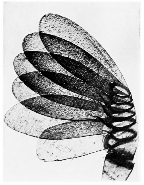
By the courtesy of Messrs. F. Davidson & Co.
The Feeler of a Cockchafer
The end of the feeler consists of a number of plates, which can be spread fanwise. The pores visible on the plates are the insect’s organs of smell.
Amongst the Wracks, of which there are a number of kinds, the methods of increase reach a higher stage. First of all let us describe the plants, so that we may know what to look for. They all belong to the group of brown Algæ. The “Channelled Wrack” is, when fully grown, about six inches long. It is much branched, often almost yellowish in colour and grows just below high-water mark. Along one side of the plant there is a moderately deep groove. Here we may note that the Wracks grow in zones from just below high water mark to low water mark. A little nearer the sea than the haunts of the Channelled Wrack, we shall find the Flat and Bladder Wracks. The former is but six inches or so in length, with flat, forked fronds, along the centres of which runs a single rib. The Bladder Wrack varies considerably in size. It may be smaller than either of the Wracks we have already mentioned or it may be two or three feet in length. It is the one seaweed familiar to everybody. Nearer to low tide mark we shall encounter the Knobbed[234] Wrack, greenish brown in colour and often as much as six feet in length. It is so named because from the sides of its flat, leathery, strap-like fronds, there arise little stalked bladders. Right at, and often beyond, low tide mark there dwells the Notched Wrack; very similar to, though larger than, the Flat Wrack, from which it may easily be distinguished by the fact that the edges of its fronds are toothed, after the manner of a saw.
It is obvious that the structure of any one of these Wracks is much more complicated than is the case with Ectocarpus. The latter Alga was composed of a number of cells, similar to one another in every respect except size. If we tease a stem or a frond of one of the Wracks upon a slide and examine the result of our efforts under the microscope we shall see that the cells which compose the Wrack are not by any means similar to one another. Those of us who have mastered the, by no means difficult, art of section cutting, should cut sections of stem and frond and compare them with sections of leaf and stem of some higher plant. The comparison will show us that, although the seaweed does not exhibit the complicated structure of a flowering plant, it has at least three kinds of different cells—an outer layer, a central structure and an intermediate layer.
If we secure a specimen of the Channelled Wrack, at the end of the summer, we shall notice that the tips of certain of the fronds are swollen. Examination of these swellings under a low magnification[235] will reveal a number of wart-like structures, and at the end of each wart there is a little pore. If we open up one of these little warts, very carefully with our mounted needles, we shall find that each little pore opens into a cavity, within which we can find two kinds of structures, hidden amongst a number of hair like growths. We shall see a number of dark, oval bodies, at the base of the hairs, these are the egg cells; more careful search will show us a number of much branched structures also partly concealed by the hairs, these are the male organs of the plants. The purpose of the hairs, by the way, is to keep the little chamber moist, when the plant is left high and dry. If we watch the pores of the other warts carefully we may be fortunate enough to see the process of increase taking place, for it occurs outside the plant and not within the chamber. The egg cell divides into two and its contents pass out of the chamber by way of the pore; each cell of the male organs gives rise to sixty-four oval little structures, each provided with a pair of minute whip-like threads by means of which it swims from the chamber and goes in search of the egg cells. Many of these little navigators are lost by the way but one of them will reach and fuse with each egg cell. After fusion the new-formed cell germinates at once into a new Channelled Wrack.
That this is an advance is shown by the fact that the little swimming bodies which fail to fuse with the egg cell, do not develop into new plants as in Ectocarpus nor does the egg[236] cell, which has failed to fuse with a swimming body, germinate.
In the Bladder Wrack, a very similar process takes place. There are, however, certain important differences, differences which show that the plant is still more highly developed. If we examine the cavities, in the little warts of the Bladder Wrack we shall find that some of them contain egg cells, some contain male organs but none contain both. We noticed that the egg cell of the Channelled Wrack produced two eggs, that of the Bladder Wrack produces eight. In other respects the two plants behave similarly.
The methods of increase amongst the red seaweeds are rather more complicated and as our object is to interest and not to puzzle our readers we will content ourselves with a few general remarks. Microscopists who are anxious to probe more deeply into the subject will soon devise ways and means for themselves. The little swimming bodies which lend an added attraction to the study of the brown seaweeds are replaced, amongst the red Algæ, by organisms with but one whip-like structure apiece and that without the power of propelling its owner through the water. As with Ectocarpus, increase may take place in two ways. On these red plants we may find the now familiar swellings, which we have learned to know are spore cases, but instead of the multitudes of free swimming organisms which are set free on the bursting of the brown sea weed spore case, we now witness the expulsion of but four[237] inert spores, which settle down in the water and immediately grow into new plants.
In the second method of increase, where male and female organs are concerned, we find that both these structures grow on the outside of the plant and not in cavities. Let us take the common, pink, much branched seaweed, known by the fearsome name of Callithamnion Corymbosum as our example. The male organs grow in little fungus like tufts about the branches of the plant and they give off enormous numbers of little organisms which have no power of swimming to the female organs. Either on the same or on another plant we shall find the female organs; we need not describe them in detail but there is one point of very great interest. From each of the female organs there grows a long jelly-like hair. As we have remarked, the organisms set free by the male organs cannot swim about but float aimlessly in the water. Obviously the majority of them simply perish, one perchance may touch a sticky hair to which it adheres, with which it fuses and passes down to the female cell, resulting in the production of a new seaweed.
It may be remembered that in writing about the pollen grains of flowering plants, we mentioned that those plants dependent upon wind for the distribution of their pollen, have stigmas ingeniously contrived for catching and retaining the grains. It is curious that the red seaweeds should have very similar contrivances for capturing and retaining the male cells.
The sea will also provide us with a rich harvest of those beautiful microscopic objects, the Diatoms. They may be sought on seaweeds, their yellowish brown colour often betrays their whereabouts, on rocks and sand and in mud. The salt water forms are as varied and as beautiful as their fresh water cousins.
To the microscopist who merely uses his microscope for the pleasure he can derive from it, rather than for serious study, it may appear that the plant life of the sea falls short of the animal life as far as interest is concerned. He may disabuse himself at once of this idea. There is no class of plants more interesting than the seaweeds and in few branches of plant life is there greater scope for new discoveries. The garden of the sea is largely an unexplored territory and there is no coast-line in the world of equal extent which provides so many different sea dwelling plants as our own.
Those of our readers who have borne with us thus far may quite excusably have thought that the last word had been attained in the construction of the microscope. It is true that different makers have made various improvements to their instruments, from time to time in recent years, most of them of minor importance but useful in the aggregate. But a few years ago, however, the advent of the Micro-Telescope and Super-Microscope marked an epoch in the manufacture of the microscope. We have shown that great strides were made in scientific investigation when the first simple lenses were manufactured, that there was a lull in microscopic research till the appearance of the compound microscope and now, when the latest instruments are in the hands of scientific workers, further possibilities are opened up.
In all microscopes—the best as well as the cheaper instruments—there is one failing which very early forces itself upon the user. They have very little[240] “depth of focus.” Let us explain exactly what the phrase means. Once or twice in our pages, we have recommended that the fine adjustment should be rotated to and fro while certain objects are being examined. When potato starch grains, for instance, are magnified sufficiently highly to show their characteristic markings, the whole of the grain cannot be seen clearly at one time, because at that magnification the “depth of focus” is slight. The higher the magnification the less is the “depth of focus”; when this quality is absent altogether only one plane of an object can be viewed clearly without re-adjusting the focus. With low magnifications, we may, to a limited extent, have more than one plane of an object in focus.
The same question of “depth of focus” occurs in photography and perhaps an example showing how it affects the camera user may make the matter clearer. Suppose we wish to photograph a landscape having, let us say, a tree in the foreground, a cottage in the mid-distance and a hill in the distance. If our lens is one of large aperture, that is to say admits a considerable amount of light and is also what is known as a long focus lens we shall find, when we view the scene on the ground glass, that when the tree is sharply focussed, the cottage and hill are not clear. When we rack in the camera to get the cottage sharply focussed, the tree and hill will be un-sharp. Similarly when we focus on the hill the tree and cottage remain out of focus. The reason is that the lens in our case possesses[241] little “depth of focus.” The experienced landscape photographer, did he wish all three objects to appear equally sharp could easily attain his object. He would perform the simple operation known as stopping down his lens, that is to say he would gradually close its diaphragm, while viewing the scene on the ground glass. At a certain point everything would be sharp, from foreground to distance. At the smaller aperture of the lens, caused by closing the diaphragm, the depth of focus would be considerably increased, at the same time much less light would be admitted to the camera. Looking upon our object, under the microscope, as comparable to a landscape, seen on the ground glass focussing screen of a camera, it is obvious that, unless our object has no thickness, and this is impossible, we cannot highly magnify its upper and lower surfaces at one and the same time. There are no adjustable diaphragms in the objective so our only course is to examine the two surfaces in turn or to resort to a lower magnification.
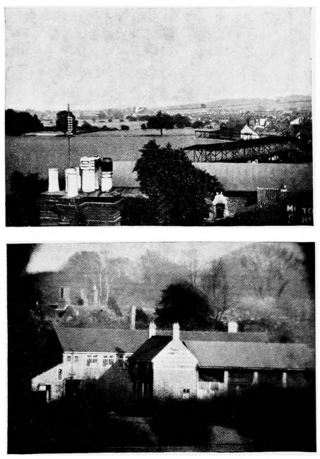
By the courtesy of Messrs. F. Davidson & Co.
The Micro-Telescope
1. View taken with ordinary camera. The arrow shows the building illustrated in the plate below; it is 3/4 mile away.
2. The building indicated by the arrow in the plate above, taken from the same standpoint through the micro-telescope.
Apart from any other consideration, the super microscope marks a big advance from the fact that it possesses great “depth of focus.” It is possible, for example, with this remarkable instrument to examine a moss as it grows, with a high magnification and see not a portion of a leaf or a fragment of the stalk, as with the ordinary microscope, but the whole upstanding plant, in stereoscopic relief. It shows us objects exactly as we should see them were we endowed with super eyes, enormously enlarged,[242] in relief and erect. Objects viewed through this instrument are not inverted, as with the ordinary microscope.
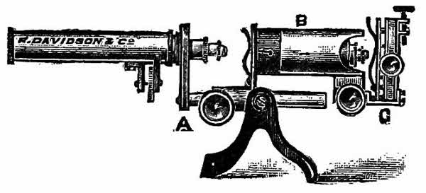
A is the microscope proper and is, in all respects, similar to the instrument with which we are familiar, except that its mirror and condenser have been removed. In the fitting provided for the condenser a second microscope is arranged.
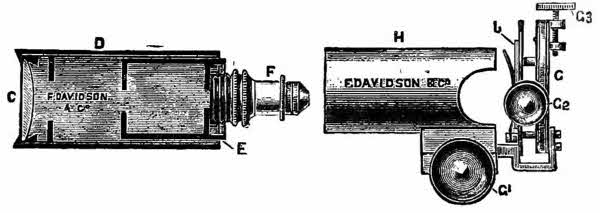
This consists of a tube D, an objective F, an ocular C and an inner sliding tube E, the whole fits into the metal case B, G is a stage on which the object is placed and below G, if necessary, a condenser may be fitted.
The instrument owes its remarkable magnifying powers to the fact that the additional microscope B, forms a magnified image of the object on the stage G, at the opening in the stage of the microscope A. This magnified image is still further[243] enlarged by the original microscope. In other words, the power of one microscope is employed on that of another.
The apparatus as we have described it gives enormous magnifications and it is possible, by using a suitable combination of objectives, to obtain a magnification of ten thousand diameters. Expressed in non-technical language, a circle whose actual diameter is equal to the thickness of the paper upon which these words are printed, could be so magnified by the super-microscope that it would appear to be as thick as ten thousand similar pieces of paper but, with this enormous magnification, it is obvious that, even with such a marvellous instrument, the whole of the circle from edge to edge could not be seen at one time. With an ordinary microscope a magnification of one thousand three hundred diameters is considered highly satisfactory. So that there is a great probability of being able to see some of the minute objects which are known to exist, but which have, up to the present, eluded those who would view them, on account of their minuteness. There is a possibility also of discovering a new underworld of which no man has yet dreamed. The man who uses his microscope solely because of the pleasure he derives from it, rather than he who uses it because it is essential to his business or profession, will be more attracted by the micro-telescope. By this we do not infer that the instrument is not useful, as a fact it is of the greatest importance in certain cases, especially[244] for Nature-study work, for the observation of minerals and to the chemist.
Like its sister instrument, the micro-telescope consists of an ordinary microscope to which is attached a specially designed object glass in a tube, which take the place of the condenser. The makers, indeed, provide two of these object glasses—one for viewing objects from one to two feet away, the other for viewing objects from a distance of three feet to the planets, if need be. Once when the instrument was being tested some crumbs were placed on the floor at a distance of four yards and strongly illuminated, and the microscope with a 1-inch objective focussed on the crumbs. With this objective in an ordinary microscope the magnification would be about thirty diameters. Presently some mice came out, and made themselves at home with the crumbs. The mice could be examined at this distance, without their being aware of it, so well that individual hairs were easily visible and about half a mouse was in the field of view. In point of size each mouse appeared about the same as a beaver within a foot or two.
Messrs F. Davidson & Co., 29 Great Portland Street, London, are well known for the high state of perfection to which they have brought the micro-telescope and other instruments connected with microscopy.
The novelty of the micro-telescope will appeal strongly to the Nature lover. At a distance of a few feet a spider can be magnified to the size of a large[245] cat, and it can be watched spinning its web with spinnerettes the size of teacups. Ants at a distance of six feet are seen to be fearsome individuals, six inches in length, and their tiny burdens are so magnified that they appear like yule logs or goodly-proportioned boulders, according to their nature. At a distance of ten feet a wasp may be seen scraping tiny shavings of wood from oak palings by means of its jaws—shavings which it converts into paper for building its nest. Very small insects may be observed as they come into the world from their chrysalis stage. The never-tiring jaws of the caterpillar may be seen at work devouring some favourite leaf—the whole action of biting and swallowing the vegetable matter can be plainly seen. Such interesting events as the tending of green flies by ants, the leaf-cutting habits of the leaf-cutter bee, and a hundred and one other events are all revealed by the micro-telescope and at such a distance that the living objects are not disturbed in their activities, being quite unaware that they are under observation. To the botanist the instrument is no less useful.
In various manufactures, such as ore smelting, and in the manufacture of glass, china and pottery, in enamelling and in certain engineering shops, where it is necessary to examine material at a high temperature, the micro-telescope is a great boon, and, at least, is the means of avoiding considerable physical discomfort. It is used also by architects and surveyors for examining the condition of the factory[246] chimneys, bridges, derricks and the like. To the engineer the instrument is invaluable; we have explained elsewhere how necessary it is that various metals and mixtures of metals should be examined for fractures. Under the high magnification with the ordinary microscope, even with an instrument specially designed for the work, only a very small area can be studied at once. With the micro-telescope a relatively large area may be examined under a high magnifying power.
There are three features of this ingenious apparatus which cannot fail to commend themselves equally to the casual worker and the serious microscopist. All objects are seen erect, just as the eye sees them. This is brought about because, as in the case of the super-microscope, we really observe a magnified, inverted image of our object, formed at the spot where the object would be placed were we using an ordinary microscope. This image is inverted once more in the microscope and so it appears to us erect.
All objects appear in relief and in their proper planes; this is seen in a striking manner by viewing an ordinary photograph through the apparatus, when the various parts stand out as in nature. There is also enormous depth of focus. With the attachment for viewing objects at a short distance, the whole of a tubular shaped flower such as a daffodil may be in focus at once, from the tip of the petals to the bottom of the tube. With the attachment designed for long distance work, objects[247] from twenty yards to sixty miles may be clearly viewed at one and the same time.
A few of the unsolved problems confronting the microscopist are reviewed in our concluding chapter. Whether they will ever be solved we dare not venture to say; but, if they are to be, surely to this latest arrival in the microscopic world and to its companion, the super-microscope, the honours will go.
To thoroughly comprehend the various uses to which the chemist may put his microscope, it is necessary to have a knowledge of chemistry. The science is so wide in its scope that no single chapter could do justice to it. There are analytical chemists, scientists whose aim is to find out the composition of various substances; biological chemists who deal with the many problems of life in which chemistry plays a part, but we need not attempt to detail all the branches of this highly specialised science.
Chemical analysis is founded upon the fact that when certain chemicals are mixed together they will, or they may, unite to form quite a different chemical. This newly formed chemical is probably a different colour to the substances which were used in its making, or again the original chemicals may be soluble in water and the new chemical insoluble, in which case it will form a cloudiness known as a precipitate. An example may help to make our[249] description clearer. Suppose we take some common table salt and dissolve it in a little water in a glass, then add to this a little solution of nitrate of silver, which is sold under the name of lunar caustic, we shall find that a white cloudiness is formed when the liquids mix, although originally they were both clear. The reason of this cloudiness is that the two substances, dissolved in water have united with one another to form a third substance which will not dissolve, therefore it settles down as a fine powder. Long experience has taught analytical chemists exactly what chemicals to add to test for all the common substances, by the formation of these precipitates; so that, if any powder were given to one of these scientists he could tell its composition by applying certain tests. In this chemical analysis considerable quantities are required and it is often necessary to test very small samples, so small that the ordinary methods are out of the question. This is where the microscope scores, because with this wonderful instrument, only drops are required and tests of corresponding delicacy may be made; in fact, by modern microscopic methods it is possible to detect the presence of as little as ten thousandth part of a grain of arsenic, or quicksilver or of the deadly poisons, Strychnine or Prussic Acid.
In testing for poisons the microscope is invaluable. Frequently only the most minute traces of the poison occur in the system and the modern microscopist who makes a study of poisons and their[250] detection can solve mysteries which would have baffled all the scientists in the world in days gone by.
Our chemical studies with the microscope may well begin with various common crystals; they are usually easy to prepare; the process of crystallization is always interesting to watch, and as objects for the microscope it would be difficult to find anything more beautiful than these home-made gems. All crystals should be examined by reflected as well as by transmitted light. When we are working with the former lighting, a piece of black paper beneath the slide will help to show up the objects to better advantage.
The easiest method of obtaining crystals of any substance which is soluble in water is to make a saturated solution in this liquid, to put a small drop upon a slide, then to tilt the microscope slightly so that there is a thin film of solution at the upper side of the drop. The microscope must not be tilted so much that the liquid runs from the slide on to the stage. Where the film of liquid is thin crystals will be found first. A word of explanation is necessary concerning the term saturated solution, especially as we may have occasion to use it many times. When we add a solid to a liquid in which it is soluble, we shall find that the liquid will take up a certain amount of the solid and no more; when, on the addition of more solid it fails to dissolve, we have reached the saturation point. A saturated solution then is[251] a liquid in which the maximum amount of solid is dissolved.
We have already described the most simple way in which crystals may be formed, and we may easily make objects for our microscope in this manner of all the substances we can find which are soluble in water. With some we shall find that crystals do not form easily, in which event we may modify our tactics and warm one drop of saturated solution on the slide till nearly all the moisture is driven off, then there should be no difficulty in watching the crystals in process of formation. Common salt, sugar, alum, borax, washing soda, iron sulphate, called also green vitriol and copper sulphate, or blue vitriol, are common and easily obtained substances, all soluble in water. As we carry our experiments a little further we shall find that crystals formed from cold solutions as suggested in our first method differ from those formed from hot solutions. Again, if we use some other substance than water as our solvent the crystals which separate out will differ once more. Many very interesting experiments may be tried on these lines.
Many beautiful crystals may be obtained by dissolving various substances in gelatine or gum. The method is simple. Gently warm a little gelatine, to which is added an equal volume of water, in a chemist’s test-tube. When the gelatine has all dissolved make a saturated solution of the substance, from which crystals are derived. Green or blue vitriol are good subjects for the experiment. Add a little of[252] the saturated solution to the gelatine and stir with a glass rod, taking care to avoid the formation of air bubbles. A little of the mixture may now be placed in a thin film on a slide covered with a cover slip and left to cool. Examination when cool under the microscope will show lovely fern-like crystals, whose beauty rivals that of ice crystals familiar to us on our window panes during hard weather in the winter. Barium chloride also produces very beautiful fern-like crystals when treated in this manner. Chlorate of potash, familiar to most of us as a remedy for sore throat, forms crystals totally dissimilar to those substances we have named. Having made as many crystals as we wish from water and also from gelatine solutions, we may turn our attention to gum arabic. The method of obtaining crystals from this substance is exactly similar to that described for gelatine except, of course, that we substitute gum for gelatine. This work is of the greatest interest, for not only does it yield wonderfully beautiful objects for our microscope, but it is a hobby full of surprises. When we are about to examine a new crystal for the first time we can never so much as hazard what shape it will assume. Sugar, it may be mentioned, does not crystallize at once from a saturated solution in water, and the best method of obtaining the crystals is to warm the slide, on which we have placed a drop of solution, and then when dry to set aside for a day; at the end of that time, especially if the air be moist, the crystals will have formed.
We shall find it interesting to try experiments in mixing two different substances, then we shall probably obtain crystals totally unlike those of either of the ingredients. As an example of this method, let us make a saturated solution of a mixture of blue vitriol and magnesium sulphate in water. Place a drop of the solution on a slide, heat over a flame, not only till dry, but till the substance left on the slide begins to melt. We must use every care not to crack the slide, and this may best be accomplished by keeping it moving while over the flame. Now if we watch our object through our microscope we can witness the formation of wonderful feathery crystals, as the slide cools.
Some strikingly beautiful results may be obtained in another manner; the method is used by analysts in their so-called fusion tests. We take a small grain of some substance, say, lead nitrate, place it in the centre of our slide, cover with a cover slip, and warm over a flame till it melts. Then, taking a similar-sized grain of another substance, such as saltpetre, we place it against the edge of the cover slip on the side opposite to the lead nitrate. Further warming will cause the saltpetre to melt, and run below the slide and mix with the lead nitrate. If we watch the meeting of the two chemicals a wonderful sight will reward us. The lead compound forms a “beautiful crystalline skeleton,” whilst the saltpetre forms six-sided stars at the opposite side of the cover slip. The experiment may be repeated, using lunar caustic and saltpetre, also the iodides of silver and[254] potassium; in fact we may try any chemical substances we have at hand, though we shall find that some do not melt very easily, and potassium chlorate is somewhat dangerous, for it forms explosive compounds with certain substances.
The number of interesting and beautiful objects which may be obtained by so-called solution tests is practically unlimited, and the enthusiastic microscopist will certainly try all the tests we describe as well as many others of his own invention. Should the chemist wish to detect the presence of aluminium in very small quantities, he relies on his microscope and proceeds in the following manner. He takes a drop of the solution, suspected of containing alum, and adds to it a drop of sulphuric acid. This mixture he puts on his slide, which he heats over a flame till dry. Now he adds a drop of water, and then a very small amount of calcium chloride is brought to the edge of the water. Beneath his microscope he can now watch the formation of beautiful colourless, eight-sided crystals which denote the presence of aluminium.
A word of warning is necessary concerning this and the following experiments; they may not always succeed, for success largely depends upon having the solutions at the right strength, and experience alone can teach us what is correct. Interesting and easily formed crystals may be obtained from barium sulphate, and the experiment may also be made to show the phenomenon which we have already mentioned, that the form of the[255] crystals depends on the nature of their solution. A little barium sulphate should be dissolved in strong sulphuric acid. Here, by the way, another warning: every care should be taken in the use of all acids; they should never be allowed to come into contact with face, hands or clothes, nor should they touch any part of the microscope. Some acids give off fumes, and these should not be allowed to reach the eyes or nose, and the microscope must be protected from them. To continue our experiment: While hot a drop of the solution of barium sulphate in sulphuric acid should be transferred by means of a glass rod to a slide and allowed to cool. Examination with the microscope will show that the barium sulphate has formed small rectangular scales. With the remainder of the sulphuric acid we now make a saturated solution of barium sulphate, and find, on repeating the method described above, that the chemical has formed curious x-shaped crystals. Similar experiments may be tried with calcium sulphate with the certainty of interesting results.
Many crystals of calcium, in the form of calcium oxalate, may be found in plants, and they are well worth looking for. They may best be seen in sections of the plants, but, if we have not mastered the art of cutting sections, we may find them by teasing the plant cells apart with our mounted needles. In the stems of rhubarb there is the substance in bundles of long needle-shaped crystals, to which the name of raphides has been given. In the seed of the garden poppy, just below the skin, there is a layer[256] known as the crystal layer, where crystals of calcium oxalate occur as tiny balls called crystal sand. In the leaf stalks of begonias very beautiful and occasionally very large crystals of this substance may be found, whilst in shapes they are strikingly varied. In orris root there are enormous crystals of calcium oxalate; in fact it is common in many plants.
If we have a photographic friend who will supply us with quite a small quantity of gold chloride, we shall be in a position to try three most interesting experiments and to obtain some curious crystals. We require a very weak solution of gold chloride in water, not more than 3.5 per cent. for our first experiment. Mix one drop of this solution with the same quantity of hydrochloric acid on a slide and heat over a flame till dry. The microscope will now show us probably the most curious crystals we have ever examined; some are long, some short, and some a zigzag in form; mixed with these there will be a few flat plate-like crystals with rectangular projections. All these curious crystals are yellow.
If instead of hydrochloric acid we use a solution of common salt in water and repeat the experiment as before, we shall obtain pale yellow prisms and some crystals of common salt. Gold is costly, so it is perhaps lucky that one of the tests for this rare metal is one of the most delicate known to chemists; it is possible, in fact, to detect very minute quantities of gold. For this experiment we may take an exceedingly weak solution of gold chloride and place a drop on our slide; we also require a solution of the[257] chloride of tin, known as stannous chloride, in an evil smelling liquid called chlorine water. If now we watch our drop of gold solution under the microscope, and while watching mix with it a drop of stannous chloride solution, a strikingly beautiful purple colouration is produced—this purple has been named the purple of Cassius.
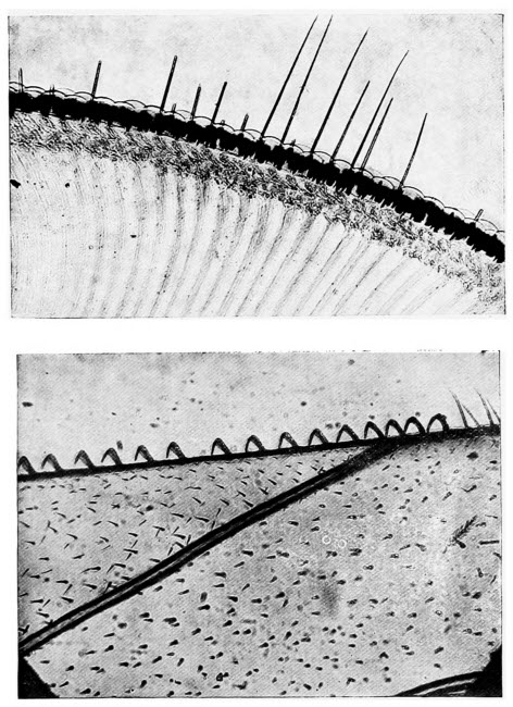
By the courtesy of Messrs. F. Davidson & Co.
1. The Eye of a Cockchafer
This section shows the eyelashes, the convex lenses, and, in the lower portion of the plate, the nerve fibres leading from the brain to the eye.
2. Hooks on Bee’s Wing
The row of hooks on the margin of the bee’s wing are clearly shown. By their aid the fore and hind wings are fastened together when the insect flies.
If we desire further experiments in the testing of common substances, and incidentally in the production of beautiful crystals, we might do worse than try the effect of adding a solution of platinum chloride to any solution containing a compound of potassium. Charmingly beautiful crystals will result.
The experiments we have described as well as hundreds of others are used by analysts every day in the testing of various substances. We have started in every case by knowing what our solutions contain; the duty of the analyst is to discover what he has before him. Given an unlimited quantity of a substance for testing purposes it is not always easy to determine its composition. With very small quantities, perhaps less than a tea-spoon full in all, the difficulties of the analyst are increased tenfold and without the assistance of the microscope his efforts would be unavailing, he deals in drops and every drop is precious. Sad to relate this form of testing, known to science as micro-chemical analysis, has been practised to a far greater extent on the continent than in this country.
Those of our readers who wish to try the experiments[258] we have described for themselves can obtain all the necessary substances, except the poisons, at any chemists and the quantities required will only be the smallest that can be obtained, in fact any reasonable-minded chemist would probably let a microscopist whom he knew have a few grains of a large number of chemical substances, suitable for this work, at the outlay of a few pence. The chlorides of gold and platinum we fear no one will give away.
All the experiments we have described thus far have necessitated the use of what are called inorganic substances, they may be described in every day language as substances derived from the inanimate world. There are many equally interesting tests which may be carried out with animal and vegetable products.
Formic acid is the substance which renders the sting of ants so painful; it may, however, be prepared artificially and if a little, dissolved in water, is mixed with a solution of silver nitrate we shall obtain flat plate like crystals also some resembling fine fibres.
Probably the most curious of the easily obtained crystals from vegetable products, may be made from citric acid which occurs in lemons. If a little of the acid be mixed with a solution of caustic soda and boiled with calcium chloride, a drop of the liquid after boiling placed on a slide will give crystals readily. When viewed from above they are an elongated oval shape, described by some[259] authorities as resembling a whetstone. Viewed from the side they have a striking similarity to small sheaves of wheat.
With the recognition of poisons under the microscope we need not trouble ourselves here. It would be useless to describe any of the experiments, for few of us could obtain such deadly substances as nicotine, strychnine, aconite, morphia and the like. Nevertheless the recognition of these and similar substances in very minute quantities is rendered easy, to those who have the necessary knowledge, by means of the microscope.
In how many branches of commerce, we wonder, does the microscope play its part. It is used in several departments of engineering for examining steels and many other metals not only for defects but to see how they are made up. It is used in brewing for studying the various yeasts and other substances, including the hops which go to the making of beer. All manufactures which depend upon fermentation, such as wine and vinegar making, are largely dependent upon the work of the microscope. In dairy work the microscope is invaluable. In the examination of various fabrics the assistance of the microscope is always summoned. Paper manufacture and paper testing give work for the microscopist but it would, we think, be easier to give a list of the branches of commerce in which the microscope is not used than to attempt to enumerate those which make use of the instrument.
We cannot possibly describe all the uses to which[261] the microscope is put, so we will confine ourselves to one or two of the more important and, at the same time, to those which can, for the most part be repeated at home.
The two most important commodities for mankind are food and clothing; we cannot live without food and those of us who take but little pride in our appearance, must have clothing of some sort. We have said a little about food in another chapter and there we have also mentioned the impurities which find their way, by accident or design, into some of the commoner foods.
In this chapter we will deal first of all with clothing describing how many of the raw materials may be recognised under the microscope and showing very briefly how fraud in connection with the manufacture of wearing apparel is detected. Practically all clothing is made from animal or vegetable fibres, some, however, is made of artificial fibres and these we shall mention.
The vegetable fibres used in the manufacture of wearing apparel are all either hairs or what are called bast fibres and the latter, in non-scientific language, may be described as the strands which run through the roots and stems of most plants. The chief requirements of vegetable fibres, destined to be woven into fabrics, are strength, it is obvious that a weak fibre would be useless; length, the longer the fibre the better and as we shall see later, short fibres are often made up into inferior material; pliability, a stiff fibre would make an[262] uncomfortable fabric; firmness and durability. Animal fibres used in the manufacture are either hairs or silk.
The most important vegetable fibre is cotton, it consists of the hairs from the seed coats of several species of Gossypium, a plant closely related to our common mallow. There are very many different kinds of cotton and the qualities of the fibres of these different cottons vary tremendously. Each hair is one cell and more or less spindle shaped, that is to say, thicker towards the middle than at the base. If we can obtain a little raw cotton we should certainly examine it under the microscope; this may best be done by laying one or two fibres in a drop of water on a slide. Under the low power, the first thing that will attract our attention is the fact that the fibres are twisted, corkscrew fashion, though not regularly nor throughout their whole length. This curious twisting makes raw cotton easily recognised and it is, at the same time, a very valuable peculiarity of these plant hairs. The greater the number of twists and the greater their regularity, the more valuable the cotton becomes for weaving purposes. Under a higher magnification, we recognise other characteristics of the cotton fibre. Each fibre is somewhat flattened, its edges are thick and, running up the centre, there is a fairly broad lumen, as it is called. Covering the whole there is a skin which by the way is often wanting in the fibres of cotton fabric owing to the chemicals with which the[263] raw cotton has been treated and also to the methods of manufacture.
A very striking experiment may be tried by soaking a few cotton fibres in cuprammonia, a substance prepared by the action of ammonia solution on copper filings. Constrictions occur at fairly regular intervals along the fibre so that, after treatment with cuprammonia, the cotton fibres resemble strings of little beads.
The manufacture of mercerised cotton has become very important of late years. The process is named after its inventor, Mercer, and consists in removing the skin from the fibres, causing them to untwist and, by doing so, to impart to them a lustre of silk. We may make a little mercerised cotton for examination under the microscope by soaking some raw fibres for a short time in a solution of caustic soda or caustic potash and then washing them in water to which a little acid has been added. This will cause the fibres to untwist and also destroy the skin, but we shall probably notice that the fibres have shrunk. In the process of manufacture precautions are taken to prevent this shrinking for then the lustre is much better. We shall also observe that in our mercerised fibres the lumen has become very narrow and it is often broken, here and there are swellings on the outside of each fibre, corresponding to the positions of the twists. Mercerised cotton, in addition to its lustre, is stronger and absorbs dyes more easily than ordinary cotton.
Flax consists of the bast fibres of the flax plant.[264] Examination of the raw product under the microscope will reveal both long and short fibres. The former are the more valuable and are used in the manufacture of linen, the latter are made into tow. The long fibres, which are derived from the upper parts of the flax plant have thickened edges and a very small lumen. The short fibres, used for making tow, come from the lower part of the stem and the roots of the plant. Each fibre has a broad lumen and is very similar to hemp fibre. Examination of all these fibres, by the way, is best made in water as described under cotton.
Hemp is another bast fibre and as we have remarked it resembles the short fibres of flax; there is a broad lumen with an indistinct margin. If we have an opportunity of comparing these fibres under the microscope we shall see that many of those of hemp have forked ends. This is very characteristic of the plant and is never found in flax, therefore it affords a ready means of distinguishing hemp from flax. Fine linen should never contain hemp, so that if our object be to test the quality of a sample of linen by microscopic examination, we must keep a sharp look out for the forked fibres of hemp. In coarse linen these fibres occur for hemp is used in its manufacture.
Jute, another important fibre is readily distinguished under the microscope, for its margins have perfectly smooth walls and its lumen is wide in some places, narrow in others and interrupted altogether in places.
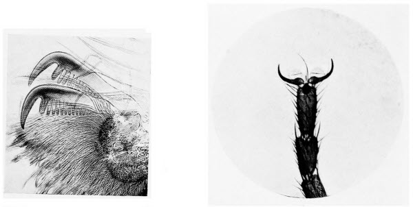
Photos by Flatters & Garnett
A Spider’s Foot
The toothed claws are well adapted to enable their owner to obtain a firm grasp of the fine threads of its web.
The Foot of a Fly
The two claws enable the fly to walk up rough surfaces, whilst the suckers between the claws give it a firm hold on smooth surfaces.
There are an extraordinary number of vegetable fibres which are woven into articles of commerce, of one kind and another. Then again, many fibres are so short or so brittle that they cannot be woven but are used for other purposes such as filling cushions, cheap bedding, etc. There are also a certain number of vegetable fibres which are valuable because they are stiff and bristle-like as well as durable, and they are used for brushes, door mats and for similar purposes. To the microscopist who is interested in this work there is a wide field open.
For the examination of paper, which may be described as a “felt of finely divided fibres,” the microscope is invaluable. The essentials of a good paper are that it be durable, that it retain its colour and not become brittle. The least observant of us cannot fail to have noticed that there are an extraordinary number of different kinds of paper, not only the many kinds which the paper manufacturers could show us, but the obviously varied papers which we meet with every day. Added to the papers, there are cardboards which are really a kind of paper. It is clear, therefore, that the man who can tell us exactly how any and every paper is made and what it is made of has laid up a goodly store of knowledge. In carrying out tests of paper we rely partly on chemical and partly on microscopic tests.
A number of substances contribute to the manufacture of paper; linen and cotton rags, hemp and[266] various fibres are the most commonly used, not forgetting wood pulp which we shall mention in a moment. The finest and whitest paper is made from linen rags and that from unused linen and hemp is the strongest. Without attempting to describe in detail or even in outline the different processes which the various vegetable fibres must undergo before they appear in the guise of paper, we may say the treatment is very drastic. Strong chemicals and machinery designed to reduce the fibre to the finest possible particles render the examination of paper, for the purpose of discovering its composition, far from easy. Such fibres as survive the rough treatment are mere fragments yet they are often large enough for the lynx eyed microscope to read their story. Formerly the constituents of most papers could be separated into three classes according to their behaviour with iodine solution, but this test has been superseded by more complicated methods which do not concern us here.
The examination of various papers may prove interesting for example in linen rag paper, we ought to find some flax fibres, they will be sadly battered and torn but are usually recognisable under the microscope. Hemp paper is tough and is used for bank notes, in it some of the short tow fibres will probably occur and they will give a clue to its composition. Cotton rag paper is easily recognised for the fibres are very characteristic, a remark which also applies to jute paper, the so-called manila used for envelopes, wrappers, etc.
Mechanical wood pulp which enters so largely into the manufacture of paper is easily recognised by those who have given a fair amount of attention to the microscopical examination of plant life. Wood pulp is always used in conjunction with some binding material such as cotton or flax fibres. Many different kinds of wood are converted into pulp and of course it requires a considerable amount of experience to say exactly what kind of wood has been used in a certain paper. Some of the woods are poplars of various kinds, others spruces and firs. It is easy to distinguish the conifers as spruces and firs are called, for the reason that the trees bear cones. The little fragments of wood, scattered throughout the paper, have minute circular perforations upon them, resembling miniature quoits, if they belong to the conifers; none of the other woods possess these “pits” as they are called.
Very many other plant remains may be found in paper, for instance hop fibres are used sometimes and their presence is usually shown by remnants of the climbing hooks which, during life, studded the climbing stem of the hop.
Some of the important animal hairs, used in the manufacture of clothing, may now claim our attention. Wool and silk are of course the most important. The best wool is all obtained from the domestic sheep so let us examine one or two of the easily obtained hairs from this animal. As with the vegetable fibres we may examine them in a drop of water but, in this case, we shall find that we[268] cannot make the water go near the wool for the reason that the latter is covered with a film of grease, called wool fat. Our first care, therefore, it to get rid of the wool fat and this may be done by shaking the specimen with a little ether or chloroform.
Having cleaned the wool fibres we may proceed to examine them under a high magnification and, to see their structure fully, we must move the fine adjustment to and fro, for it is not possible to obtain a true idea of its structure without doing so. We shall see that the hair consists of two layers, an outer skin and an inner core. The latter consists of a number of cells, whilst the former is composed of scales, of which the lower edges are arranged beneath the upper edges of the previous scales, like tiles on a roof. The free edges of the scales project outwards a little so that the wool, when not so highly magnified, appears to have a toothed margin. Further examination will show us that no single scale completely envelops the strand of wool, usually two scales make the complete circuit. It is a curious and easily tested phenomenon that in say an inch of wool from the same kind of sheep there are always the same number or very nearly the same number of scales. By counting the scales, experts can tell from what animal the wool is derived.
If we examine many samples of wool we shall not be long before we encounter certain specimens showing one or more constrictions. Now we all[269] know that the reason why some people do not grow very much is because they are delicate, ill health affects the whole system. The constrictions in the sheep’s wool occur because the animal has suffered from some illness, or from great hunger or thirst, which has resulted in its wool not growing properly for a period corresponding to the duration of the illness or other calamity.
The examination of various animal hairs will help to while away many an hour and many of these objects are of the greatest interest. If we have the opportunity it will be interesting to compare the wool from different kinds of sheep, that from the Lincoln sheep, for example differs from that of shortwooled kinds. We may also compare goat’s wool with sheep’s, then there are differences between the hair of cows and calves. Comparisons always make microscopic work more interesting.
The microscopic examination of cloth, used for making our coats, is quite interesting work and withal important. A good deal of the cloth which is made up into suits is known as shoddy, that is to say that material that has been worn before; old rags of all sorts and many other extraordinary things go to the making of this cloth. There are special factories to which rags are sent to be made up into shoddy. One might think therefore that this substance would be easily recognised under the microscope but it is not quite so easy as we shall see. Absolutely pure wool from the sheep giving the best quality wool is only used in the very finest[270] and most expensive fabrics. In a great deal of really good cloth we may recognise other hairs besides those of the sheep and sometimes vegetable fibres find their way into good cloth by accident. A piece of suspected cloth should be cut off and separated as far as possible into its different fibres. In shoddy we shall find few long fibres and they will all be much torn for the reason that the rags from which the material is made are cut up in the manufacture. If we find cotton fibres, we may be certain that our specimen is shoddy, also if we can find fibres of many different colours, though the final dyeing may have disguised the fact that the fibres have originally figured in fabrics of various colours.
Silk is one of the most important of all fibres capable of being woven into fabric. It is hardly necessary to remark that it is formed by the fully fed silkworm just before it turns into a chrysalis. A very large number of caterpillars spin silk but the majority of this silk is useless for commercial purposes. The silkworm gives off a double thread of silk from glands in its mouth and, at the same time, it gives off a sticky substance called silk glue which sticks the two fibres together, so that, to the eye at least they appear as one fibre.
Most of us have kept silkworms, those who have not may find it worth while to expend a few pence in some of these insects for the sake of examining the raw silk. A cocoon, as the work of the caterpillar[271] is called, consists of three layers, an outer layer of floss, a middle layer and a so-called layer (the inner layer) of parchment. Only the middle layer is used in commerce, the floss is too fine and weak and the parchment is so impregnated with silk glue as to be useless.
If we examine some raw silk, taken from the middle layer of a cocoon, we can easily see the two parallel fibres of silk and the outer wrinkled covering of silk glue. Now, magnifying our object more highly, we shall see that each fibre is a solid rod, with a smooth lustrous surface and without any sign of lumen or cell structure; the rods too are continuous and this alone distinguishes silk from all other fibres animal or vegetable. Two tests are worth trying for they are characteristic of silk. On the addition of a little strong sulphuric acid we observe that the silk rapidly dissolves, on the other hand, if a few fibres are boiled in hydrochloric acid, the silk dissolves but the envelope of silk glue remains unchanged and appears beneath the microscope as a cracked and wrinkled tube.
The caterpillar of another moth spins coarser greyish coloured fibres, which are spun into the well known Tussore silk. As with the common silkworm these caterpillars spin two fibres and glue them together with silk glue. In this case, however, under a high magnification we shall notice that the fibres are marked with a number of very fine lines running lengthways, whilst every now and then there are fairly deep indentations. The[272] former markings are natural to the fibres, the latter are caused by one fibre being pressed against the other. In countries where the production of silk is of great importance, the microscope is not only pressed into service for examining the product of these useful little insects, but also in keeping watch for a very deadly disease which attacks the caterpillars. It is called “pebrine” and the great scientist Pasteur, whose name is world famous for his work on bacteriology, discovered that it was caused by a tiny fungus.
Artificial silk is an important article of commerce. It is made in several different ways and of various substances. Some artificial silk is made of collodion, some again is made of cellulose the substance of which the cell walls of young plants is composed; gelatine is also used in making this commodity. As a rule it is easy to distinguish artificial from real silk, for usually the imitation consists of flat fibres or at least fibres quite different to the smooth rods of real silk. The iodine test is often sufficient indication, for with this chemical true silk is coloured brown.
Space or lack of it does not allow us to describe how the microscope may be and is applied to other manufactures, even the miller uses the instrument, for it will tell him if his flour is of pure wheat, and in this manner. He puts a little flour in a drop of water on a slide and covers with a cover slip; then, for a moment or two, he rubs the cover glass to and fro over the water and flour and examines his[273] specimen under the microscope. If his flour be of wheat, he will see fairly stout spindle shaped strings, if it be of rye no strings appear, whilst maize flour gives very small strings. These are called gluten strings and wheat is very rich in gluten.
Photography is such a popular hobby in these days, that the enthusiast who possesses both camera and microscope, is certain, sooner or later, to wish to take permanent records of some of the beautiful objects revealed to him by the latter instrument. The production of high-power photo-micrographs, as the pictures of highly magnified objects are called, can only be carried out by those who are skilled in the use of both camera and microscope and are possessed of considerable patience. There is nothing to prevent any amateur photographer who possesses a bellows camera—box cameras cannot be used for this work—from producing excellent low-power photo-micrographs, that is to say pictures of objects less highly magnified.
It may be disappointing to learn that a microscope is not necessary for this work. The requirements are, a camera which will open out to a considerable extent and a short focus lens. Before we show how the pictures may be taken, we must[275] be quite clear what is meant by a short focus lens. There are various ways of measuring the focus of a lens; in the case of a single lens the operation is fairly simple, but single lenses are made of one glass and most camera lenses are built up of several pieces of glass, then it is much more difficult to measure the focus quite accurately. For ordinary purposes and for our purpose, it is only necessary to know the focus in round figures and to do so we open up the camera and focus some distant object on the ground glass, then the distance in inches from the back of the lens to the ground glass will give us very nearly the correct focus, or as it is often called focal length, of our lens. Suppose we focus on a church steeple a mile away and then find that a space of five inches separates the back of our lens from the ground glass, the lens is of five inches focal length.
With a lens of such a focal length we shall require a camera with very long bellows to obtain much magnification of our object but, if our camera is one with only short bellows, we can still overcome our difficulties. If the extra expense is no object we can obtain one of the excellent Aldis lenses of only two inches focal length, especially designed for this work; if we are ingenious we can construct a device which will answer our purpose admirably and cost but a few pence. With the simple apparatus we are about to describe photo-micrographs up to ten diameters magnification can be obtained in most cameras. The term, ten diameters,[276] may appear puzzling but really it is quite simple. Suppose we wish to make a photo-micrograph of a penny stamp and we wish it to be magnified ten diameters, we should require a large camera for the operation, but that, for the moment, is beside the point. Our penny stamp, magnified ten diameters, would be equal in area to ten horizontal rows of stamps, each row containing ten stamps or, in other words, it would occupy the same area as one hundred penny stamps.
To find the magnification possible with our camera and lens, extend the camera bellows to the full and set up a foot rule in front of the lens. Move the camera from a distance slowly towards the rule till it is sharply focussed, then carefully measure the distance between the inch lines on the focussing screen; if the lines are three inches apart we shall be able to make photo-micrographs three times the size of our object and we shall probably desire something better.
To obtain a considerably magnified picture we must have a lens of short focal length and a camera with long bellows, in fact in theory the amount of magnification is only limited by the length of the bellows, so that an extraordinarily long camera should give us a much magnified picture. We cannot lengthen our bellows without considerable expense, but we can shorten the focal length of our lens. If we obtain, at the cost of a few pence, a convex lens of short focal length, such as is used in cheap magnifiers we can easily[277] apply it to our camera lens and it will have the effect of shortening its focus. This extra lens can either be fitted in front of our original lens by cutting two inches of cardboard, of such a size that they will just fit into the lens hood. In the centre of each piece of cardboard, cut two circles, slightly smaller in diameter than the diameter of the convex lens. Place the lens over the opening in one of the pieces of cardboard and stick the other piece upon it with glue or seccotine. When dry fix the lens in its cardboard holder in the front of the lens hood and repeat the focussing experiment with the foot rule. We shall now obtain a much greater magnification with the same length of bellows, because the additional lens has shortened the focus of our original lens. Probably our camera is fitted with a double lens and it is possible to unscrew the front portion, in this event our extra lens with its cardboard holder may be fitted inside the front lens up against the diaphragm and the front lens replaced. The result is practically the same whatever the position of the new lens, but we must be certain that it is convex, for a concave lens would increase the focal length of the whole and so reduce our magnification.
We may find that the amount of enlargement we can now obtain is sufficient for our purpose; if so we can go ahead and produce photo-micrographs of all the objects we desire. The methods of doing so differ in no way from those employed in taking ordinary photographs and as we are writing about[278] microscopy and not photography in these pages, there is no need to go into further details. Even the experienced photographer, however, is liable to overlook one or two important details. The bugbear in all work of this kind, with low magnifications as well as high, is vibration. Not only is our object magnified but every movement is equally increased. No one should walk across the room while this work is in progress; in large towns, trams and heavy motors will ruin many a plate, in fact it is only when one takes up work of this kind that one realises that one’s house is in a perpetual quiver. Some enthusiasts work at the dead of night, others suspend their apparatus on springs and invent all kinds of ingenious devices to overcome these miniature earthquakes.
If time is no object, we have an easy means of still further increasing the magnification. To do so we cut a thin sheet of copper in such a manner that it just fits our lens tube, in front of the diaphragm. Then in the very centre of the copper disc we make a hole with a “number one” needle, the hole is about one twentieth of an inch in diameter. Replace the convex lens in its cardboard holder and screw on the front portion of the camera lens. A trial will show that we have considerably increased the magnification but decreased the amount of light admitted by the lens, therefore we shall probably require an exposure as long as an hour. How about the bugbear vibration during such a long exposure is a natural question[279] to ask. Unless the vibration is continuous, and that is unlikely, it is less likely to cause trouble during a very long exposure than during a short one, because it operates during a small proportion of the whole time.
All kinds of objects can be depicted with the apparatus we have described. Minute shells and insects; parts of larger insects, their legs and wings for example; the feathers of birds; various rocks, crystals of all kinds; small flowers and their seeds; mosses, lichens and many kinds of fungus, in fact their number is limited only by the degree of ingenuity possessed by the photographer. Do not suppose that these low-power photo-micrographs are interesting only because of their size, the enthusiast who makes a collection will discover in his prints hidden beauty of which he had no conception when he looked at the originals. We have seen a very large number of low-power photo-micrographs, taken with very inexpensive apparatus, showing the wonderful sculpturing on the wing cases of beetles, some of them are marvels of design, yet, observed with the naked eye, many of the insects appear to be devoid of ornamentation.
The production of high-power photo-micrographs is hardly a subject that can be described in these pages; the apparatus is costly for, even if one dispenses with a specially designed micro-photographic camera, it is necessary to have a good microscope and bellows camera, for really advanced work. Messrs Swift & Sons supply an excellent fitting, with[280] which quite satisfactory photo-micrographs may be taken. It consists of a light metal cone, the more pointed end fits over the upper portion of the microscope tube; the other end of the cone is provided with ground and plain glass focussing screens and a dark slide. When using this apparatus, it is first necessary to find and focus our object in the ordinary way, before attaching the photographic apparatus. Having secured our object exactly as we wish it to be depicted and well in the centre of the illuminated circle, we remove the eyepiece and slip the metal camera over the top of the microscope tube. If now we place the ground glass screen in position, we can see an image of our object upon it. Great attention must be paid to the lighting, it is necessary that the illumination be perfectly even, otherwise our negative will be over-exposed in some parts and under-exposed in others. When everything is in order the plain glass screen is substituted for the one of ground glass. On this we shall probably not see any image with the naked eye, but with the help of a magnifying glass we can see a much finer image than was possible on the ground glass screen. The final focussing is done at this stage and, having secured as sharp an image as possible, the focussing screen is removed and the exposure is made. Experience alone will teach the length of exposure, it depends upon the amount and nature of the light, upon the transparency of the object and also upon its colour. There is no more simple apparatus for taking[281] moderately high-power photo-micrographs; for more advanced work the micro-telescope and the super-microscope (see Chapter XVII) used with a camera, will enable the user to obtain pictures of objects magnified as much as five thousand times.
Having described all the ordinary uses of the microscope and having also insisted that the objectives are the most important part of the instrument, there are probably many of our readers who may wish to know in what manner this wonderful glass differs from ordinary glass and how it is made.
Glass has been defined as a substance which, during its manufacture, passes from the liquid to the solid state so rapidly that no crystals are formed. Usually when solids are melted and then allowed to cool, they do so with the formation of crystals, this may be shown in the case of sulphur by melting a little and then allowing it to cool. After a while, as cooling takes place a solid crust will be formed on the surface of the molten sulphur. If two holes are pierced in the crust and the still liquid sulphur poured out, it will be found that the sulphur which adhered to the vessel in which the melting took place has formed beautiful needle shaped crystals.
A great amount of original work has been done[283] on the subject of glass manufacture and especially on the kind used for optical instruments—as a result there are many kinds of glass differing from one another in physical properties and in chemical composition. Although the various chemicals used and their proportions are fairly well standardised, as the result of long experience, it is probable that glass is not a definite chemical compound but a mixture, in which certain of the components act as solvents for the rest.
The ordinary glass in use in this country, apart from specially prepared optical glass, may be either English flint glass, plate glass or Bohemian glass. The first named is composed of sand, potassium carbonate and red lead; plate glass is made of sand with the carbonates of sodium and calcium, whilst similar ingredients are used for Bohemian glass except that carbonate of potassium is substituted for carbonate of sodium. It is chiefly owing to the requirements of optical instrument makers that, new kinds of glass containing very many previously untried chemicals, have been produced. As a result glasses are now made with specific gravities varying from 2.5 to 5.0, that is to say the weight of a square inch, or a square foot or a square yard of glass may weigh anything from two and a half to five times more than a square inch, foot or yard of water, according to its composition.
Before we describe the details of its manufacture let us consider its properties as briefly as possible. At high temperatures it is perfectly fluid and may[284] be poured from vessel to vessel as easily as water; at lower temperatures it is viscous, i.e., semi-fluid and can be rolled with an iron roller as dough is rolled with a rolling pin; it can be moulded into any desired shape, blown out into flasks and bottles or drawn out into threads so fine that they may be woven into a fabric. It is a bad conductor of heat and for this reason it is safer to pour very hot liquids into a thin glass vessel than into a thick one. With thick glass the inner layers expand with the heat before the outer layers are even warm and the result is a crack or often absolute fracture. Sometimes during manufacture glass vessels which are suddenly cooled will appear satisfactory, but the particles of glass remain in so high a state of tension that at the slightest touch the vessel will break up into thousands of pieces. On account of this property of glass it must be cooled very slowly indeed; the process is known as annealing.
Optical glass unlike most other kinds must be manufactured in thick blocks—some of the large lenses on telescopes are of considerable thickness. All glass for scientific instruments must also be homogeneous, which our dictionary tells us means of the same kind. To be more explicit each particle of optical glass should be precisely the same in composition and properties as every other particle. In the very early days of manufacture it was difficult to obtain homogeneous pieces of glass and Guinard, in the 18th century, conceived the idea of[285] stirring the molten glass with a rod of fireclay, to ensure a thorough mixing of the components. This led to considerable improvement and the method has survived to the present day.
The first real advance in the manufacture of optical glass, was due to the ingenuity of two Germans, Abbe and Schott, who lived at Jena. Jena glass became famous for the manufacture of lenses, so much so that a stupid idea still prevails in many quarters that only the Germans can make good optical glass. To give them their due it is good but quite recent events have shown the world that the Britisher can make better. The two German scientists used new chemicals in making their glass and they succeeded in producing a substance which possessed hitherto unheard of properties. In what is now known as the older crown and flint glass the dispersion and refraction increased with the density, that is to say, the heavier the glass the more it scatters and bends light rays passing through it. With the new methods, glass is made which scatters the light rays very little, though bending them considerably and vice versa. The ordinary crown glass is composed of silicates of calcium and sodium or of calcium and potassium or a mixture of both and it is possible to make it colourless and free from defects, but its optical properties are never so valuable as those of Jena glass. The most important components of the newer glass are the oxides of Barium, Magnesium, Aluminium, Zinc and Boron.
Good optical glass should be transparent and colourless and, as we have stated, it should be homogeneous—the refraction and dispersion of light rays should be identical over all parts of the glass. It should possess no striæ, as they are called. Striæ may be seen at the edge of a piece of plate glass as little lines just as though the glass had been formed in layers. Striæ detract from the efficiency of optical glass, nevertheless, some very cheap lenses are made of plate glass. Bubbles are almost always present in Jena glass but, unless they are very numerous they do not appear to render the glass less efficient. Hardness and chemical stability are other desirable qualities. Most of these high-grade glasses are soft, as shown by the ease with which they may be scratched; many of them are not very stable chemically and are easily affected by chemical fumes with the result that their surfaces become covered with a coloured film. With all their drawbacks, for optical work the newer glasses far excel the older.
The manufacture of optical glass is a costly and lengthy process. The chemicals used in its manufacture are selected with the greatest care; impurities must be guarded against for they would change the composition of the glass and in doing so alter its physical properties on which everything depends. The chemical substances are used either in the form of oxides, nitrates or carbonates, for the reason that they are easily decomposed by heat. To assist in the melting of the substances a few[287] fragments of glass of similar composition are added. The crucibles, in which the chemical components are melted, are covered so that no fumes from the furnace may gain access to them, even the chemical composition of the crucibles is carefully tested that no impurities may contaminate the glass. No crucible is ever used more than once and only a single crucible is heated in each furnace, in order that the temperature may be regulated to a nicety.
The actual manufacture is then begun after all these preliminaries have been attended to. A clean dry crucible is heated in a furnace—not the one in which the glass making is to take place—to a dull red heat. Then, with iron tongs, it is removed to the previously heated glass making furnace and the temperature is raised very gradually. The next stage sees the addition of the well mixed chemicals to the heated crucible, in small quantities at a time. When the full quantity has been added the crucible contains melted glass full of bubbles, some of them air bubbles released from the raw materials as they were added and some bubbles of gas given off from the chemicals as they act upon one another. The molten glass is then heated strongly so that it will become perfectly liquid and many of the bubbles will be driven off. To reach this stage may occupy anything from thirty-six to sixty hours and constant attention is necessary during the whole time.
The next stage is perhaps the most important in[288] the whole process. After numerous small samples of the molten glass have been taken, on the end of iron rods, to see if the air bubbles have been driven off, the mixture is stirred to render it homogeneous and to eradicate striæ. The stirring is carried out by means of a cylinder of fireclay which is first of all heated to the temperature of the molten glass before it is introduced. To the end of the fireclay cylinder a long, detachable iron handle is fixed so that the man who undertakes the stirring may stand at a distance from the hot furnace. The heat is great and the work of stirring is laborious and for this reason the stirrers are constantly changed. The iron handles must be watched carefully for, owing to the heat they rust rapidly and should any of the rust fall into the molten glass it would impart to it a colour and render it useless for optical purposes. When stirring begins the glass is liquid as water, but the stirring is continued during cooling and all the while the glass is gradually becoming more and more solid. During the final stages the operation is hard labour indeed and, finally, it is not possible to stir any more, then the fireclay cylinder is either removed or left in the glass.
When the glass has solidified and, whilst it is still hot the furnace is sealed and allowed to cool very gradually till it has reached the ordinary temperature of its surroundings; this may take several weeks. When quite cool, the crucible is removed from the furnace and carefully broken;[289] then the glass, which may be in one mass weighing as much as 1000 lbs., is freed from particles of fireclay and examined for defects.
The next stage consists of moulding and annealing. Large pieces of glass are heated till they are just soft, then they are passed into iron or fireclay moulds designed so that the glass is formed into discs or slabs suitable for grinding by opticians. They are allowed to cool very slowly in the moulds.
When the glass is taken from the moulds it is not yet ready to be made into lenses. It is subjected to another and very careful examination, when all defective parts are cut out. Should there be many defects the glass is again heated, moulded and annealed. From a crucible containing 1000 lbs. of molten glass it is unusual to obtain much more than 200 lbs. of optically perfect glass, it is obvious therefore that lenses cannot be cheap.
One would naturally imagine that the minute lenses used in microscope objectives should be cheaper in comparison than the larger lenses used for photography or in telescopes, for it is always more difficult to make minute articles than large ones. As a matter of fact, even allowing for the greater amount of material used in the larger lenses, quality being the same in both cases, they are far more expensive than the smaller microscope lenses; the reason being that it is exceedingly difficult to obtain a perfect specimen of large size. The difficulty arises not only in the actual manufacture of the glass but in the subsequent operations of[290] cutting, grinding and polishing when fractures are very liable to occur.
From this brief account of the manufacture of optical glass it is clear that a good lens is always worth a high price. Its components must be of the purest quality obtainable, the process of manufacture requires highly skilled labour and it is laborious and exacting work. Constant attention is needed from start to finish and much of the glass is never sufficiently good to pass the rigorous tests which it must undergo. Lastly, in the final preparations of the completed lens, mishaps are frequent. Added to all these trials of the lens maker is the one outstanding fact that the process can never be hurried at any stage, the efficient annealing of optical glass is one of the most important stages in its manufacture. A very full account of the manufacture of optical glass is given in the Encyclopædia Britannica, whence much of our information in this chapter is derived.
In this, our concluding chapter, we propose to give a few hints upon the choice and use of the microscope and its accessories, to enlighten our readers concerning stains and staining and to add such other information as is likely to be useful, information which is better supplied in a chapter of its own than scattered about our pages and probably overlooked.
The most important question concerns the choice of a microscope. In a book of this nature it is obviously impossible to recommend the wares of any one maker. Some firms are celebrated for the good wearing qualities of their instruments, some for the delicacy—not fragility by the way—of their fine adjustments and our readers must select their maker for themselves. One thing we strongly advise, be patriotic and select a British maker, it is not only a question of patriotism but of self interest, for in many respects the British instruments lead the world. Most of the manufacturers[292] advertise in such papers as Nature, they all print excellent catalogues, a perusal of which may be a useful preliminary.
The instrument we select will depend on the use to which we intend to put it and on the length of our purse. Perhaps the question of cost will be more important to most people. An elaborate instrument is not a desirable acquisition, till we are somewhat advanced in our work at anyrate, and one of the models which most makers call Students’ Microscopes will do everything we desire. We should have at least two objectives, three if we can afford them, a 1 inch and a 1/6 inch will enable us to examine everything described in these pages, except some of the bacteria. If we wish to add a third objective we might select a 2 inch one, for quite low-power work and later we shall probably become the proud possessors of a 1/12 inch objective, but this would be of little use to us at first. Two eyepieces will complete our optical equipment. A condenser for the stage, properly known as a substage condenser is an addition which we shall appreciate, though quite interesting work may be done without it.
Our other apparatus is inexpensive and comprises, one or two dissecting knives, some needles mounted in handles, a pair of fine scissors, a pair of small forceps, a razor, one or two camel hair brushes, three or four watch glasses, slides and cover slips, and a pipette or two. A pipette, by the way, is merely a glass tube pointed at one end and cut[293] off square at the other; at about its centre it is much wider than at the top or bottom. It is a most useful piece of apparatus for picking up small water animals which we wish to examine. By putting the pointed end of the pipette just over an Amœba, for example, and sucking gently at the other end, we shall draw some water and the animal into the swollen part of the tube. If we then place one finger over the end of the pipette to which we have applied suction we can transfer the contents wherever we wish without it running out, as soon as we remove our finger the pipette empties itself. The dissecting knives are useful for cutting up specimens before examination, the mounted needles for teasing them, that is to say tearing them into fine shreds. The brushes we may use instead of the pipette, for picking up small objects from water, or when dry. The watch glasses are useful for examining objects such as sponges, water fleas, etc., under water. They are just as satisfactory for most purposes as the specially constructed zoophyte troughs and, of course, very much cheaper. The razor is required for section cutting concerning which we will say a few words.
For section cutting, special razors are sold, they are heavy as a rule and not hollow ground. Many microscopists aver that good sections cannot be cut with a hollow ground razor, we beg to differ, however. On this point we would advise our readers to select whichever pattern suits them best; there is no need to buy an expensive razor, for our early[294] efforts, at anyrate, will quickly dull the cutting edge and there is no advantage in going to much expense in this direction.
To cut really good sections is not difficult, it is a question of knack rather than skill; having once acquired the knack, practice will do the rest. The great point is to begin properly, some people never take the necessary trouble in their early days and, as a consequence, they never learn to cut good sections.
There is nothing mysterious about the operation, a section is merely a very thin slice. A good section for examination under the microscope should, of course, be so thin that it is transparent; it should be equally thin everywhere, not thick in some places and thin in others and it should be cut in the proper direction. The last statement requires a little explanation; suppose we wish to make a section straight across a stem, a transverse section it would be called, then we must see to it that our section is cut straight across, and not in such a manner that the portion from which the section is cut tapers in the slightest degree. Before we begin the actual section cutting we must always trim up our specimen with one of the dissecting knives and not attempt to make the surface level with the razor. As a start, let us take some moderately fleshy plant stem for we shall find it as easy to cut as anything. Our stem must not be woody, for till we have had a fair amount of practice we shall damage our razor on its hard structures; it[295] must not be too soft or it will be crushed out of all recognition in our fingers; it should not be hollow, for solid stems are always easier to cut. Having selected our stem and the spot upon it at which we require a section, we cut straight across it at that spot with our dissecting knife, in such a manner that the cut edge is as nearly as possible at right angles to the sides of the stem. A piece two or three inches long is the most comfortable to hold.
Holding our razor firmly in the right hand we take our specimen in the thumb and index finger of the left hand. Place the side of the stem against the first joint of the index finger and slightly bend the tip of the finger round the object. The end of the stem, from which we propose to cut our section, should be slightly above the level of the index finger. With the thumb, the stem is pressed firmly against the first joint of the index finger and the first joint of the thumb must not be bent upwards. This last point is very important, as we shall see in a moment. From our description the method of holding the stem may seem somewhat cramped; it should not be so, however, and an easy position will help towards success but let it be the correct position. We may play the violin, in a manner, without learning the correct method of bowing but we can never become expert violinists unless we hold our bow properly, in the same way we can never cut really fine sections unless we hold our specimen in the correct way. We now lay our razor[296] along the second joint of the index finger in such a manner that the heel of the razor, or a point near the heel comes gainst the end of the stem. For the first time we realize the importance of holding our specimen correctly, for the index finger forms a convenient platform on which to steady the razor. To cut our section, we, of course, press the razor towards us and, at the same time draw it outwards from heel to toe. We must never attempt to press the razor straight through the specimen, however soft the latter may be, we must always draw the razor along, at the same time as we press it through the specimen. Never cut outwards as in sharpening a pencil and never move the razor backwards and forwards as though using a saw. The outward cut will never result in a good section, a sawing movement will give a section alternately thick and thin. Should the section be somewhat tough we may find the razor slip suddenly over its surface, an event which will impress itself upon us painfully if we have forgotten the injunction not to bend the thumb, for the certain result of the slip will be the loss of a goodly portion of skin upon one’s joint. A straight thumb will be out of the danger zone of a slipping razor.
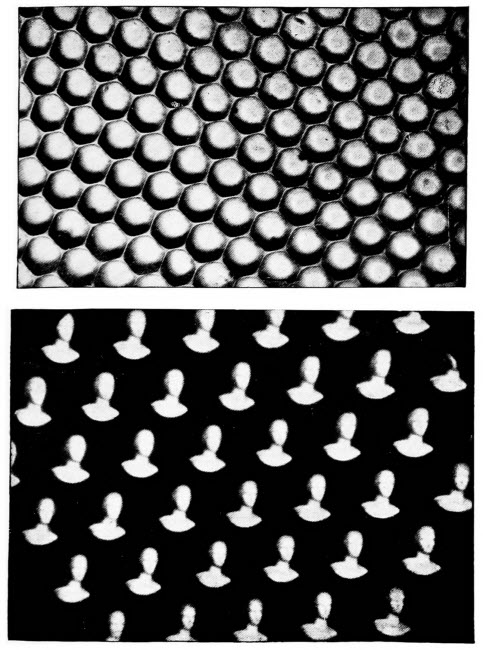
By the courtesy of Messrs. F. Davidson & Co.
1. A Fly’s Eye
A fly’s eye highly magnified, showing some of the many lenses of which it is composed.
2. Images Seen by a Fly
Photographs of a statue taken through a fly’s eye. Instead of seeing one image of an object, the fly sees as many images as there are lenses in its eyes.
Having cut a section it is hardly likely that, at this our first attempt, it will be good enough to put under the microscope, so we will continue to cut section after section till we have a dozen or more from these we can select the most transparent for examination. The sections of such material as a[297] stem should be kept moist and to do so we will place them in a watch glass containing water. It is often easier, also, to cut our sections if the razor be moistened with water, at anyrate the moisture prevents the sections from adhering to the razor. The sections should be removed from razor to watch glass and from watch glass to slide, by means of a brush, never by means of the fingers. The razor of course, should be well dried with a soft rag before it is put away; rust, besides being unsightly, ruins the cutting edge. If we are really anxious to cut our own sections, and every good microscopist does so, we shall return to the operation again and again, even cutting objects which we have no desire to examine, for the sake of the practice. We shall soon reach a stage where our razor will be dulled and require stropping and the efficient stropping of a razor is, to many people, a more difficult operation than the cutting of sections.
We may point out here, that all sections are not quite so easy to cut as the one we have taken as our example. Some objects are so soft that they need hardening with chemicals before they can be cut, some are so hard that to attempt to cut them would ruin the edge of the razor, though it is wonderful what hard substances may be cut when we have had a little experience; some are so delicate that they must needs be buried in melted wax, then object and wax are cut together and later the wax is separated from the section.
Leaves and very small stems may be sectioned[298] by the beginner, as easily as larger stems. For such objects little sticks of pith are sold as holders. Having cut of our piece of leaf from which we wish to derive a section, we make a slit, with our dissecting knife, down the middle of one end of a piece of pith. The piece of leaf is then placed in the slit and by holding the pith at the sides our piece of leaf is held firmly. Sections are cut through pith and leaf, and the two are floated in water, when the thin slices of pith will float away from the leaf sections.
Having cut a satisfactory section, let us proceed to describe the method of making a slide thereof. We will suppose that we do not wish to make a permanent preparation but one for temporary use. A clean slide must be selected, all through our pages we emphasise the cleanliness of slides, at or near its centre we put a drop of water and, lifting it with a brush, we place our section in the drop of water. If our examination is to be with a low magnification, we need not use a cover slip, nevertheless it is worth while to cultivate the habit of using one. The cover slip is not only a protection for our object but for our objective. Water you may argue cannot harm the lenses of the objective. Perhaps not, we will not argue the point but, when the water dries on the objective, it leaves a certain amount of deposit on the glass and this deposit must be rubbed off. The less often the lenses are rubbed the better for them, glass especially very highly polished optical glass, is far more easily scratched than many[299] people imagine and a scratched lens is an inefficient lens. When using high magnifications a cover glass should always cover our object and the same remark applies to objects examined in Canada Balsam. This substance is likely to cause serious trouble if it finds its way on to an objective. It must be removed, that is obvious, but it sets hard, it must not be scraped away for fear of damaging the optical glass and, as it is used to cement the lenses together, there is the great danger that any solvent used to remove the Balsam from the face of the objectives, may also dissolve their setting. Our digression may seem somewhat unnecessary, but the very great importance of keeping all chemicals and even water, from coming into contact with the lenses of our instrument cannot be insisted upon too strongly.
In whatever substance we examine our object, water, glycerine or Balsam, there is a right and a wrong way of applying the cover slip. It must not be dropped or laid down flat upon the object, if we do this we shall certainly imprison a number of air bubbles and that must be avoided. One edge of the cover slip must be laid against the edge of the mountant, as the liquid used for mounting our object is called, then placing a needle beneath the cover slip it must be gently lowered into position. We shall now find that all air bubbles are driven out as the cover slip is lowered.
When we have cut an exceptionally good section or when we have some specially interesting object we may wish to make a permanent slide. The exact[300] method of doing so varies somewhat with the nature of our object and to describe all the methods of mounting microscopic objects permanently would require a book in itself. We will suppose that we wish to make a permanent slide of one of the sections floating in our watch glass of water. The first thing we must do is to get rid of the water with which our section by now is saturated. This may be accomplished by means of alcohol of various strengths, which we may put into three watch glasses. In the first watch glass we have half pure alcohol and half water; in the second three-quarters alcohol and one part water; in the third watch glass pure or absolute alcohol. The section is transferred on a brush to the first watch glass and left there for five minutes, then to the second watch glass for a similar time and finally to the third watch glass. The mountant may be either Canada Balsam or glycerine jelly. Should we decide on Canada Balsam, we put a small drop of the substance in the centre of a clean slide, place one section atop of it and then gently lower a cover slip upon it as already described. Then, without using any force, the cover slip is pressed down, but we must not fall into the all too common error of thinking that a thick section can be made thin by pressing upon the cover slip. If we have taken the correct amount of Canada Balsam it will just cover the area below the cover slip and no more, if we have used too much, it will flow out on all sides and perhaps on the top of the cover slip. A very[301] little experience will teach how big a drop of Balsam to use.
Should we decide to use glycerine jelly, we take a small globule of the jelly on one of our mounted needles and place it in the centre of a clean slide. Then we hold the slide a little above a lighted candle, or even a match will do, to melt the jelly. Directly it is melted the section is placed upon it, and we proceed as before. Whichever mountant we have used, the slides must be put away out of the dust, and they must be flat and not placed on edge. In a few days the mountant will set, and we need take no further precautions with the slides in which Balsam was used, but those mounted in glycerine jelly must be ringed, for the reason that glycerine absorbs moisture from the air and gradually liquifies.
The process of ringing is best performed upon a turntable, which any dealer in microscope accessories will supply. It consists of a circular brass plate which revolves about its centre and to which the slide to be ringed is affixed. A bottle of ringing asphalt and a fine paint brush are essentials. With a little of the asphalt upon our brush, we revolve the turntable and place the brush against the edge of the cover slip as it revolves, in such a manner that we paint a narrow rim of asphalt over the junction of cover slip and slide. The asphalt prevents moisture from reaching the glycerine jelly. Of course we may ring our slides without making use of a turntable, but it is not easy to paint a neat ring without mechanical assistance. For square cover slips it is[302] obvious that a turntable is useless. Specimens mounted in Canada Balsam do not need ringing, for the Balsam is unaffected by air or the moisture therein, when once it has set hard.
Several firms supply prepared microscope slides, and it is often useful to know where reliable preparations may be obtained. The slides supplied by Messrs Flatters & Garnett Ltd., 409 Oxford Road, Manchester, are models of what well-made slides should be.
It always lends greater interest to the hobby if objects are found and mounted by the microscopist himself. By going out into the fields, by the pond-side, or along the shore, in search of interesting material for examination, much will be learned of animal and plant habits which even the microscope cannot reveal. Some of us, however, have neither the opportunity to hunt for our specimens, nor the time to mount them properly, and those of us who are so situated will be glad to know where objects may be obtained.
Apparatus, useful for students of pond or sea-shore life, may be obtained from Messrs Flatters & Garnett, who always have a goodly stock of collecting jars, nets, &c. From the same firm, also, may be obtained stains and any of the limited number of chemicals required by the microscopist.
We have given a few simple directions for staining in our chapter on Bacteria. In many cases it is absolutely necessary to have recourse to stains in order to see the structure of the objects we are[303] desirous of examining; in other cases it is necessary to stain when we wish to know the nature of the various parts of our object. Suppose, for example, we wish to find out whether a plant section contains starch, we then add iodine solution, and if any parts stain deep blue we know at once that starch is present. There are other stains for other plant and animal substances; stains for woody matter, stains for fats, &c., but the art of staining is a science in itself, and would require many chapters to describe fully. Most of the objects described in this book can be studied in the natural state, but, even so, they may be rendered far more beautiful by staining.
The method we described for the staining of bacteria does not apply to such objects as plant sections, &c., and we propose to describe, as briefly as possible, how to proceed with such objects. Suppose we are examining one of the common pond plants, Spirogyra for instance, and we wish to see whether it contains starch. Our specimen is in a slide, in a drop of water, and covered by a cover slip. In the first place, we must obtain some fluffless blotting paper—the ordinary filter papers sold by all chemists are excellent—from it we must cut about half a dozen pieces, about half an inch by one inch, the exact size is not important, and they need not be measured. These we must fold in the centre, so that they can be made to stand up like an inverted V. From our bottle of iodine solution we take a drop of the liquid on the end of a glass rod and place it carefully at one edge of the cover slip, avoiding allowing any of[304] the solution to flow on to the upper surface of the slip. Now, one of the pieces of blotting paper must be placed upon the opposite side of the slide so that it will stand up; it must then be moved till it just touches the edge of the cover slip. The blotting paper will absorb the water from beneath the cover slip, and in doing so the iodine solution will be drawn along to take the place of the water. By proceeding in this manner and replacing the blotting paper with a new piece as it becomes moist, also replenishing the drop of iodine as it is used up, we act upon our object with a stronger and stronger solution, and, in Spirogyra, the object which we took as our example, we can see beautiful rosettes of starch grains, arranged at regular intervals along the green bands of chlorophyll. This method of staining may be used in most cases where we merely require a temporary stain; by reversing the process and drawing water over the object by means of blotting paper, it may be used in washing sections and parts of plants. For very small objects, such as starch grains separated from the plants in which they are formed, the method is hardly suitable, for they are liable to be drawn along in the stream of liquid and lost.
For more permanent staining processes, we must use our watch glasses, into which we pour the various liquids necessary for the operation. The precise methods of staining, the periods during which objects should remain in the staining solution, and the chemicals used for removing excessive stain vary, as may be guessed, according to circumstances. Some[305] chemicals act very quickly, and staining takes place in a few minutes; others act slowly, and with them it is necessary to subject our specimens to their action for hours or even days. Then again, it is obvious that large specimens take longer to stain than small ones, hard objects are not so readily acted upon as soft ones. Experience alone will show what is required in various cases.
Suppose, for example, we desire to stain a section in Carmalum, a mixture of Carminic Acid 1 grain, Alum 10 grains, hot distilled water 200 c.c. We take three watch glasses, in one we place a few drops of our stain, in another water, and in the third alcohol. Our section is placed in the watch glass containing the carmalum and is left there for about two minutes, then with the help of a small brush it is transferred for a similar period to the watch glass containing water, and finally it is placed in the alcohol. From the last watch glass it may be transferred to glycerine jelly on a slide and mounted as already described; Carmalum stains our section a beautiful pink.
There is a temptation to buy a large stock of stains of all hues and of varied composition. The temptation should not be allowed to get the better of us. In our early days at anyrate, we shall do far better if we use but few stains and learn to understand their peculiarities.
The most useful selection for the beginner will comprise Haematoxylin, Safranin, Eosin, Carbol-Fuchsin, Methylene Blue, and Carmalum. These should all be obtained ready for use: this will save[306] errors in compounding, and, in certain cases, will save time; some of the stains, Haematoxylin for example, are not fit for use till months after they have been mixed with the other ingredients which form the complete stain.
Frequently when examining small but lively water animals we may feel the necessity of some method of sobering them. All the little organisms, both plants and animals, which are provided with the little whip-like structures mentioned so often in our pages, are difficult to examine whilst they are in motion. There are many substances, we might add, which would kill them, but in doing so we shall nearly always find that they contract to such an extent as to lose all semblance of their natural shape, and become useless as objects for our microscope. Should our inclinations lead us in the direction of much study of these little beings, we shall do well to keep handy a small bottle of Rousselet’s solution; it is composed of 2 per cent. solution of hydrochlorate of cocaine, 3 parts; Methylated Spirit, 1 part; water, 6 parts. Any chemist will mix the solution for us. When we have occasion to examine a too lively specimen, we simply run a drop of the solution under the cover slip, as described in our remarks on staining with iodine, and the creature we are examining will abandon its frolics and conveniently remain in a fully expanded state.
Many of our readers will be anxious to carry their microscopic investigations to a more advanced stage than we have reached in our pages. Some will have[307] the advantage of a teacher, and it is a great advantage to have someone who can show rather than merely explain what should be done; others not so well placed may like to know a few really useful books which will help them in their work. “The Microscope and its Revelations,” by B. H. Dallinger, is a large and therefore costly work, but it contains a rare fund of information for the microscopist. Smaller and eminently suitable for the general worker is “Modern Microscopy,” by Messrs Cross & Cole. Those who wish to specialize in one of the sciences, such as Botany, Zoology, or Geology, will find no lack of books dealing with the subject that most appeals to them. For photographers who are also microscopists, we know of no better books than “Practical Principles of Plain Photomicrography,” by G. West, or the more advanced “Handbook of Photomicrography,” by Messrs Hind & Randles.
Neither books nor teacher will be able to reveal to any of us all the secrets of the microscope. By its means a new world is unfolded before the eyes of mankind, a world of unlimited possibilities. No man will ever see all that the microscope can show him, each day some fresh wonder is looked upon for the first time.
As a hobby, microscopy can hardly be excelled. It is a sensible hobby and, after the initial outlay, need cost us but a few pence each year. We hear someone say that he prefers an outdoor hobby, but surely the microscope, if used intelligently, will take[308] us out of doors. The search for specimens is one of the attractions of microscopy. As an instrument for the serious worker the microscope compares favourably with any other invention of mankind.
Think for a moment what we owe to the microscope in a hundred and one walks of life, and then you will realize the romance attached to the instrument, and its revelations from the days of Euclid to the present time.
FOOTNOTE:
[1] Humour is here used in its original sense, meaning moisture or a liquid.
Seeley, Service & Co LTD
Founded 1795
THE NEW ART LIBRARY
“The admirable New Art Library.”—Connoisseur.
NEW VOLUME
THE PAINTER’S METHODS & MATERIALS
By Professor A. P. Laurie, M.A., D.SC., F.R.S.E., HON. R.S.A., Professor of Chemistry to the Royal Academy. With 50 illus. 21s. nett.
LANDSCAPE. By Adrian Stokes, R.A. 15s. nett.
ETCHING. By E. S. Lumsden. 208 illus. 21s. nett.
DRAWING. By Harold Speed. 96 illus. 10s. 6d. nett.
OIL PAINTING. S. J. Solomon, R.A. 80 illus. 10s. 6d. nett.
ANATOMY. Sir Alfred D. Fripp. 159 illus. 15s. nett.
MODELLING & SCULPTURE. A. Toft. 119 illus. 15s. nett.
WATER COLOUR. A. W. Rich. 60 illus. 10s. 6d. nett.
PERSPECTIVE. R. Vicat Cole. 472 illus. 15s. nett.
ANATOMY OF TREES. R. V. Cole. 500 illus. 15s. nett.
Illustration from Speed’s “Drawing.”
A Marine Reptile.
From “Geology of To-day.”
SCIENCE OF TO-DAY SERIES
LATEST VOLUME
16. WIRELESS OF TO-DAY. By C. R. Gibson, LL.D., & W. B. Cole. 6s. nett each Vol.
Some of the Volumes.
1. ELECTRICITY OF TO-DAY. C. R. Gibson, LL.D., F.R.S.E.
2. ASTRONOMY OF TO-DAY. Cecil G. Dolmage, M.A., D.C.L., LL.D., F.R.A.S.
3. SCIENTIFIC IDEAS OF TO-DAY. C. R. Gibson, F.R.S.E.
4. BOTANY OF TO-DAY. Prof. G. F. Scott Elliot, M.A.
6. ENGINEERING OF TO-DAY. T. W. Corbin.
9. PHOTOGRAPHY OF TO-DAY. Prof. H. Chapman Jones.
10. SUBMARINE ENGINEERING. C. W. Domville-Fife.
11. GEOLOGY OF TO-DAY. Prof. J. W. Gregory, F.R.S.
12. AIRCRAFT OF TO-DAY. Maj. C. C. Turner.
14. ANIMAL INGENUITY. C. A. Ealand, M.A.
15. CHEMISTRY OF TO-DAY. P. G. Bull, M.A.
THE THINGS SEEN SERIES
Cloth, 3s. 6d. nett. Leather, 5s. nett.
“Dainty and attractive little books.”—Daily Post.
NEW VOLUMES
26. THINGS SEEN IN SWITZERLAND (In Summer). Douglas Ashby.
27. THINGS SEEN IN THE PYRENEES. Capt. Leslie Richardson.
28. THINGS SEEN IN N. WALES. W. T. Palmer.
29. THINGS SEEN AT THE TOWER OF LONDON. H. Plunket Woodgate.
ALREADY ISSUED IN THIS SERIES
THINGS SEEN IN
1. JAPAN. Clive Holland.
2. CHINA. J. R. Chitty.
3. EGYPT. E. L. Butcher.
4. HOLLAND. C. E. Roche.
5. SPAIN. G. Hartley.
6. NORTH INDIA. T. L. Pennell.
7. VENICE. Lonsdale Ragg.
9. PALESTINE. A. G. Freer.
10. OXFORD. N. J. Davidson.
11. SWEDEN. Barnes Steveni.
12. LONDON. A. H. Blake.
13. FLORENCE. E. Grierson.
14. ITALIAN LAKES. L. Ragg.
15. RIVIERA. L. Richardson.
16. NORMANDY & BRITTANY. Clive Holland.
17. CONSTANTINOPLE. A. Goodrich-Freer.
18. EDINBURGH. E. Grierson.
19. SWITZERLAND IN WINTER. C. W. Domville-Fife.
20. ENGLISH LAKES. W. T. Palmer.
21. PARIS. Clive Holland.
22. ROME. A. G. MacKinnon, M.A.
23. NORWAY. S. C. Hammer, M.A.
24. SHAKESPEARE’S COUNTRY. Clive Holland.
25. CANADA. J. E. Ray.
THE IAN HARDY SERIES
By Commander E. Hamilton Currey, R.N.
With Coloured Illustrations. Extra Crown 8vo. 5s. nett each.
Each of the volumes in the Ian Hardy Series is a complete story by itself, but each describes another stage of the adventurous life of the delightful young rascal who is their hero.
VOLS. I TO IV OF THE IAN HARDY SERIES.
IAN HARDY, NAVAL CADET.
“An ideal gift book for a boy, for it is utterly devoid of the milksop piffle in which such stories usually indulge. A whole budget of mirth and adventure.”—Manchester Courier.
IAN HARDY, MIDSHIPMAN.
“Combines as much adventure as the most spirited of young readers could possibly wish for, with a faithful picture of what a middy’s life may be when he is lively and as plucky as Ian”—Glasgow Herald.
IAN HARDY, SENIOR MIDSHIPMAN.
“It is no exaggeration to say that Commander Currey bears worthily the mantle of Kingston and Marryat.”—Manchester Courier.
IAN HARDY, FIGHTING THE MOORS.
“Commander Currey is becoming a serious rival to Kingston.”—Yorkshire Observer.
Specimen Binding.
A STIRRING STORY FOR BOYS.
JACK SCARLETT, SANDHURST CADET. By Major Alan M. Boisragon. Coloured Illustrations. 5s. nett.
“A dashing youth who has a good time, but emerges from his training with a good character.”—Manchester Courier.
THE LIBRARY OF ROMANCE
Lavishly Illustrated. Ex. Crown 8vo. 6s. nett each volume.
“Splendid volumes.”—The Outlook.
“Gift books whose value it would be difficult to over-estimate.”—The Standard.
“This series has now won a considerable and well-deserved reputation.”—The Guardian.
40. THE ROMANCE OF OUR WONDERFUL WORLD. P. J. Risdon.
Quelling a Mutiny.
From “The Romance of Piracy.”
THE ROMANCE OF—
SUBMARINE ENGINEERING. T. W. Corbin.
MODERN EXPLORATION. A. Williams, F.R.G.S.
MODERN INVENTION. A. Williams.
MODERN ENGINEERING. A. Williams.
ANIMAL WORLD. E. Selous.
M. LOCOMOTION. A. Williams.
ELECTRICITY. C. R. Gibson, LL.D., F.R.S.E.
MOD. MECHANISM. A. Williams.
INSECT LIFE. E. Selous.
MINING. A. Williams, B.A.
PLANT LIFE. Prof. Scott Elliot.
BIRD LIFE. John Lea, M.A.
PHOTOGRAPHY. C. R. Gibson.
CHEMISTRY. Prof. Philip, F.R.S.
MANUFACTURE. C. R. Gibson.
EARLY BRITISH LIFE. By Prof. Scott Elliot, M.A., B.SC.
GEOLOGY. E. S. Grew, M.A.
ANIMAL ARTS & CRAFTS. John Lea, M.A.
EARLY EXPLORATION. A. Williams, F.R.G.S.
MISSIONARY HEROISM. Dr. Lambert, M.A.
MIGHTY DEEP. A. Giberne.
POLAR EXPLORATION. G. F. Scott.
SAVAGE LIFE. Prof. S. Elliot.
SIEGES. E. Gilliat, M.A.
THE SHIP. Keble Chatterton.
ASTRONOMY. H. Macpherson.
SCIENTIFIC DISCOVERY. C. R. Gibson, LL.D., F.R.S.E.
PIRACY. Lieut. K. Chatterton.
WAR INVENTIONS. T. W. Corbin.
COMMERCE. H. O. Newland.
MICROSCOPE. C. A. Ealand, M.A.
RAILWAYS. T. W. Corbin.
COAL. C. R. Gibson, F.R.S.E.
SEA ROVERS. E. K. Chatterton, B.A.
LIGHTHOUSES & LIFEBOATS. T. W. Corbin.
SCIENCE FOR CHILDREN
Specimen Binding.
Popular Science for Young Children of 8 to 14. Each 5s. nett.
“Among writers on science for boys, easily the most skilful is Mr. Charles Gibson.”—The Nation.
NEW VOLUME
MACHINES & HOW THEY WORK. C. R. Gibson, LL.D., F.R.S.E.
EARLIER VOLUMES
PHOTOGRAPHY & ITS MYSTERIES. C. R. Gibson, F.R.S.E.
THE GREAT BALL ON WHICH WE LIVE. C. R. Gibson, F.R.S.E.
“An admirable introduction to about half a dozen of the most recondite sciences without going beyond the comprehension of any intelligent child.”—Scotsman.
OUR GOOD SLAVE ELECTRICITY. C. R. Gibson, F.R.S.E.
“Reads like a fairy tale, yet every word is true.”—Dundee Courier.
THE STARS AND THEIR MYSTERIES. C. R. Gibson, F.R.S.E.
WAR INVENTIONS AND HOW INVENTED. C. R. Gibson, F.R.S.E.
CHEMISTRY AND ITS MYSTERIES. C. R. Gibson, F.R.S.E.
GREAT INVENTIONS & HOW THEY WERE INVENTED. C. R. Gibson, F.R.S.E.
THE WONDER LIBRARY
Particularly handsome Gift Books for Young People. Illustrated. 3s. nett each.
NEW VOLUME
THE WONDERS OF MINING. A. Williams, B.A., F.R.G.S.
Binding of the Wonder Library.
SOME OF THE EARLIER 18 VOLS. ARE:—
1. THE WONDERS OF ASIATIC EXPLORATION. A. Williams, F.R.G.S.
2. THE WONDERS OF MECHANICAL INGENUITY. A. Williams, B.A.
3. THE WONDERS OF ANIMAL INGENUITY. H. Coupin, D.SC., and John Lea, M.A.
4. THE WONDERS OF THE PLANT WORLD. Prof. G. F. Scott Elliot.
5. THE WONDERS OF THE MODERN RAILWAY. T. W. Corbin.
6. THE WONDERS OF THE INSECT WORLD. Edmund Selous.
7. THE WONDERS OF MODERN ENGINEERING. A. Williams, B.A.
8. THE WONDERS OF BIRD LIFE. John Lea, M.A.
9. THE WONDERS OF MODERN CHEMISTRY. Prof. J. C. Philip, F.R.S.
10. THE WONDERS OF ELECTRICITY. C. R. Gibson.
11. THE WONDERS OF MODERN INVENTION. A. Williams, B.A.
12. THE WONDERS OF MODERN ASTRONOMY. Hector Macpherson, M.A.
13. THE WONDERS OF SAVAGE LIFE. Prof. Scott Elliot, M.A., B.SC.
18. THE WONDERS OF SCIENTIFIC DISCOVERY. C. R. Gibson, F.R.S.E.
THE DARING DEEDS LIBRARY
With many Illustrations in Colours. 5s. nett each.
9. DARING DEEDS OF GREAT BUCCANEERS. Norman J. Davidson, B.A. (Oxon.)
DARING DEEDS OF
1. FAMOUS PIRATES. Lieut. Commdr. Keble Chatterton, B.A.(Oxon.)
2. TRAPPERS AND HUNTERS. E. Young, B.SC.
3. INDIAN MUTINY. Edward Gilliat, M.A.
4. DARK FORESTS. H. W. G. Hyrst.
5. GREAT PATHFINDERS. E. Sanderson, M.A.
6. GREAT MOUNTAINEERS. Richd. Stead, B.A.
7. POLAR EXPLORERS. G. Firth Scott.
8. AMONG WILD BEASTS. H. W. G. Hyrst.
THE LIBRARY OF ADVENTURE
With Jackets in 3 colours & Illustrations. 6s. n. each.
LATEST VOLUME
11. ADVENTURES OF MISSIONARY EXPLORERS. Dr. Pennell, Grenfell, Barbrooke Grubb, Bishop Bompas, &c. R. M. A. Ibbotson.
“Of all books on Missions we have read this is the most fascinating.”—Dundee Courier.
RECENTLY ISSUED IN THIS SERIES
Adventures on the
1. HIGH MOUNTAINS. R. Stead, B.A., F.R.H.S.
2. GREAT FORESTS. H. W. G. Hyrst.
3. GREAT DESERTS. H. W. G. Hyrst.
4. GREAT RIVERS. R. Stead, B.A., F.R.H.S.
5. WILD BEASTS. H. W. G. Hyrst.
6. HIGH SEAS. R. Stead, B.A.
7. ARCTIC REGIONS. H. W. G. Hyrst.
8. RED INDIANS. H. W. G. Hyrst.
9. TRAPPERS & HUNTERS. E. Young, B.SC.
10. SOUTHERN SEAS. R. Stead, B.A.
SEELEY’S MISSIONARY LIVES FOR CHILDREN.
With Covers in 4 Colours. Illustrated. Price 1s. nett.
“With attractive coloured covers, well written, and at the wonderfully low price of one shilling. Will bring delight to the boys and girls who get them.”—Missionary Herald.
BARBROOKE GRUBB OF PARAGUAY. C. T. Bedford.
MOFFAT OF AFRICA. N. J. Davidson.
ARNOT OF AFRICA. Nigel B. M. Grahame.
BISHOP BOMPAS OF THE FROZEN NORTH. Nigel B. M. Grahame, B.A.
LIVINGSTONE OF AFRICA. C. T. Bedford.
JOHN WILLIAMS OF THE SOUTH SEA ISLANDS. N. J. Davidson.
HANNINGTON OF AFRICA. Nigel B. M. Grahame, B.A.
PENNELL OF THE INDIAN FRONTIER. Norman J. Davidson, B.A.(Oxon).
JUDSON OF BURMA. Nigel B. M. Grahame, B.A.
THE PRINCE’S LIBRARY
Handsome Gift Books for Young People. Many Illustrations. 4s. 6d. nett each.
LATEST VOLUME
17. THE COUNT OF THE SAXON SHORE. Prof. A. J. Church.
RECENTLY ISSUED
1. LAST OF THE WHITECOATS. G. I. Whitham.
2. DIANA POLWARTH, ROYALIST. J. Carter.
3. THE FALL OF ATHENS. Prof. Church, M.A.
4. THE KING’S REEVE. Edward Gilliat, M.A.
5. CABIN ON THE BEACH. M. E. Winchester.
6. THE CAPTAIN OF THE WIGHT. F. Cowper.
7. GRIMM’S FAIRY TALES. New Translation.
8. THE WOLF’S HEAD. Edward Gilliat, M.A.
9. ANDERSEN’S FAIRY TALES.
10. CAEDWALLA. Frank Cowper.
11. THE ARABIAN NIGHTS’ ENTERTAINMENTS.
12. ROBINSON CRUSOE. Daniel Defoe.
13. THE VICAR OF WAKEFIELD. Goldsmith.
14. CRANFORD. Mrs. Gaskell.
15. PATRIOT AND HERO. Prof. Church.
16. MINISTERING CHILDREN. Charlesworth.
HEROES OF THE WORLD LIBRARY
Each Volume fully Illustrated. Ex. Crown 8vo. 6s. nett.
The Storming of Jhansi.
From “Heroes of the Indian Mutiny.”
LATEST VOLUME
9. HEROES OF THE INDIAN MUTINY.
Edward Gilliat, M.A., sometime Master at Harrow School.
With Full-page Illustrations.
“A capital book.... Full of stories of heroic deeds, intrepidity, and determination performed in the face of fearful odds.”—The Field.
RECENTLY ISSUED IN THIS SERIES.
Heroes of
1. MISSIONARY ENTERPRISE. Claud Field, M.A.
2. PIONEERING. Edgar Sanderson, M.A.
3. MISSIONARY ADVENTURE. Canon Dawson, M.A.
4. MODERN CRUSADES. E. Gilliat, M.A.
5. MODERN INDIA. E. Gilliat, M.A.
6. THE ELIZABETHAN AGE. E. Gilliat, M.A.
7. MODERN AFRICA. E. Gilliat, M.A.
8. THE SCIENTIFIC WORLD. C. R. Gibson, F.R.S.E.
THE DICK VALLIANT SERIES
By Lieut.-Commander JOHN IRVING, R.N.
With Coloured Illustrations. Extra Crown 8vo. 5s. nett.
DICK VALLIANT, NAVAL CADET.
A stirring story of the life & adventures of a naval cadet in peace & war.
Dick Valliant is the first volume of a new series of books for boys which will deal with the most exciting & interesting naval incidents of the Great War. It will be followed in 1928 by the second volume entitled
DICK VALLIANT IN THE DARDARNELLES.
A new volume will be added each year thereafter.
THE PINK LIBRARY
Books for Boys & Girls. Illustrated. 2s. 6d. nett each.
NEW VOLUME
29. THE PIRATE. Capt. Marryat.
SOME OF THE 29 VOLS. IN THIS SERIES
1. LIONHEARTED. Canon E. C. Dawson.
3. TO THE LIONS. Prof. A. J. Church.
9. A GREEK GULLIVER. A. J. Church.
15. MISSIONARY HEROES IN ASIA. J. G. Lambert, M.A., D.D.
16. LITTLE WOMEN. L. M. Alcott.
17. GOOD WIVES. L. M. Alcott.
18. MISSIONARY HEROES IN AFRICA. J. G. Lambert, D.D.
21. GRIMM’S FAIRY TALES. A New Translation.
22. WHAT KATY DID AT HOME. Susan Coolidge.
28. MISSIONARY HEROINES IN INDIA. Canon Dawson.
Boxes from Home.
From “What Katy Did.”
MISSIONARY BIOGRAPHIES—a new series
With many Illustrations and a Frontispiece in Colours, 3s. 6d. nett each.
LATEST VOLUME
12. BISHOP PATTESON OF THE CANNIBAL ISLANDS. E. Grierson.
“The life story of Bishop Patteson is of such thrilling interest that boys and girls cannot help being fascinated by it.”—Teachers’ Times.

Vol. 3.
Vol. 4.

Vol. 1.
Vol. 2.
EARLIER VOLUMES
1. A HERO OF THE AFGHAN FRONTIER. The Life of Dr. T. L. Pennell, of Bannu. A. M. Pennell, M.B., B.S.(Lond.), B.SC.
2. MISSIONARY CRUSADERS. Rev. Claud Field, M.A.
3. JUDSON, THE HERO OF BURMA. Jesse Page, F.R.G.S.
4. ON TRAIL AND RAPID BY DOG-SLED AND CANOE. Bishop Bompas’s Life amongst Red Indians & Esquimo. Rev. H. A. Cody, M.A.
5. MISSIONARY KNIGHTS OF THE CROSS. Rev. J. C. Lambert, M.A., D.D.
6. MISSIONARY HEROINES OF THE CROSS. By Canon Dawson.
7. LIVINGSTONE, THE HERO OF AFRICA. By R. B. Dawson, M.A.(Oxon).
8. BY ESKIMO DOG-SLED & KAYAK. By Dr. S. K. Hutton, M.B., C.M.
9. MISSIONARY EXPLORERS. By R. M. A. Ibbotson.
10. ARNOT, A KNIGHT OF AFRICA. Rev. E. Baker.
11. BARBROOKE GRUBB, PATHFINDER. N. J. Davidson, B.A.(Oxon)
Binding of the Russell Series.
THE RUSSELL SERIES FOR BOYS & GIRLS
A Series of handsome Gift Books for Boys & Girls. With Coloured and other Illustrations. 3s. 6d. nett each.
LATEST VOLUME
12. STORIES OF EARLY EXPLORATION. By A. Williams, B.A.(Oxon.)
SOME OF THE 11 EARLIER VOLUMES
STORIES OF RED INDIAN ADVENTURE. Stirring narratives of bravery and peril. H. W. G. Hyrst.
STORIES OF ELIZABETHAN HEROES. E. Gilliat, M.A., sometime Master at Harrow.
STORIES OF POLAR ADVENTURE. H. W. G. Hyrst.
STORIES OF GREAT PIONEERS. Edgar Sanderson, M.A.
STORIES OF GREAT SCIENTISTS. C. R. Gibson, F.R.S.E.
BOOKS ON POPULAR SCIENCE
By CHARLES R. GIBSON, LL.D., F.R.S.E.
“Mr. Gibson has fairly made his mark as a populariser of scientific knowledge.”—Guardian.
“Mr. Gibson has a fine gift of exposition.”—Birmingham Post.
MODERN ELECTRICAL CONCEPTIONS. Illustrated. 12s. 6d. nett.
In the Science of To-Day Series. Illustrated. 6s. nett each.
SCIENTIFIC IDEAS OF TO-DAY. A Popular Account of the Nature of Matter, Electricity, Light, Heat, &c. &c.
ELECTRICITY OF TO-DAY.
WIRELESS OF TO-DAY. By Charles R. Gibson, F.R.S.E., & W. B. Cole.
In the Romance Library. Illustrated. 6s. nett each.
THE ROMANCE OF MODERN ELECTRICITY.
THE ROMANCE OF MODERN PHOTOGRAPHY.
THE ROMANCE OF MODERN MANUFACTURE.
THE ROMANCE OF SCIENTIFIC DISCOVERY.
THE ROMANCE OF COAL.
HEROES OF THE SCIENTIFIC WORLD. 6s. nett.
WHAT IS ELECTRICITY? Long 8vo. With 8 Illustrations. 6s. nett.
THE AMUSEMENTS SERIES
By CHARLES R. GIBSON, LL.D., F.R.S.E.
Extra Crown 8vo. Fully illustrated. 5s. nett each.

VOL. III.
CHEMICAL AMUSEMENTS & EXPERIMENTS.
NEW VOLUME. VOL. II.
SCIENTIFIC AMUSEMENTS & EXPERIMENTS.
VOL. I.
ELECTRICAL AMUSEMENTS & EXPERIMENTS.
“Dr. C. R. GIBSON’S BOOKS ON SCIENCE HAVE A WORLD-WIDE REPUTATION. Every experiment is so clearly explained and skilfully handled that the lesson is learnt at once without difficulty. He has such an engaging way of imparting his knowledge that he is entertainingly educative. An admirable gift book, packed with illustrations showing how the experiments can be carried out without much trouble or expense.”—Yorkshire Observer.
CLASSICAL STORIES. By Prof. A. J. CHURCH
“The Headmaster of Eton (Dr. the Hon. E. Lyttelton) advised his hearers, in a recent speech at the Royal Albert Institute, to read Professor A. J. Church’s ‘Stories from Homer,’ some of which, he said, he had read to Eton boys after a hard school day, and at an age when they were not in the least desirous of learning, but were anxious to go to tea. The stories were so brilliantly told, however, that those young Etonians were entranced by them, and they actually begged of him to go on, being quite prepared to sacrifice their tea-time.”
Profusely illustrated. Extra Crown 8vo. 5s. nett each.
The Children’s Æneid
The Children’s Iliad
The Children’s Odyssey
The Faery Queen & her Knights
The Crusaders
Greek Story and Song
Stories from Homer
Stories from Virgil
The Crown of Pine
Stories of the East from Herodotus
Story of the Persian War
Stories from Livy
Roman Life in the Days of Cicero
Count of Saxon Shore
The Hammer
Story of the Iliad
Story of the Odyssey
Heroes of Chivalry & Romance
Helmet and Spear
Stories of Charlemagne
THE KING’S LIBRARY
With many Coloured Illustrations. 5s. nett each.
LATEST VOLUME
7. CRANFORD. Mrs. Gaskell.
THE CHILDREN’S ODYSSEY. Prof. A. J. Church
THE CHILDREN’S ILIAD. Prof. A. J. Church
A KNIGHT ERRANT. N. J. Davidson, B.A.
THE CHILDREN’S AENEID. Prof. A. J. Church
VICAR OF WAKEFIELD. Goldsmith
THE FAERY QUEEN. Prof. A. J. Church
THE MARVEL LIBRARY
Fully Illustrated. Each Volume, 4s. nett.
LATEST VOLUME
10. MARVELS OF ANIMAL INGENUITY. By C. A. Ealand, M.A.
MARVELS OF
1. SCIENTIFIC INVENTION. By T. W. Corbin.
2. AVIATION. Maj. C. C. Turner, R.A.F.
3. GEOLOGY. By E. S. Grew, M.A.
4. WAR INVENTIONS. By T. W. Corbin.
5. PHOTOGRAPHY. C. R. Gibson.
6. THE SHIP. By Lieut-Com. E. Keble Chatterton, R.N.V.R.
7. MECHANICAL INVENTION. By T. W. Corbin.
8. WORLD’S FISHERIES. By Sidney Wright.
9. RAILWAYS. By A. Williams.
Specimen Binding.
TWO EXCELLENT RECITERS
Each volume consists of over 700 pages, cloth, 6s. nett each. Also Thin Paper Edition (measuring 6 3/4 × 3/4 ins., and only 3/4 of an inch thick), price 7s. 6d. nett each. They also contain practical Introductions by Prof. Cairns James, Professor of Elocution at the Royal College of Music.
THE GOLDEN RECITER
Selected from the works of Rudyard Kipling, R. L. Stevenson, Conan Doyle, Thomas Hardy, Austin Dobson, Christina Rossetti, Maurice Hewlett, A. W. Pinero, Sydney Grundy, &c., &c.
“An admirable collection of pieces both in prose and verse.”—Spectator.
“Offers an unusual wealth from authors of to-day.”—The Athenæum.
“Far superior to anything we have yet seen.”—Western Press.
THE GOLDEN HUMOROUS RECITER
From Anstey, Barrie, Major Drury, J. K. Jerome, Barry Pain, A. W. Pinero, Owen Seaman, G. Bernard Shaw, &c., &c.
“A most comprehensive & well-chosen collection of some hundreds of pieces. A small encyclopædia of English humour.”—The Spectator.
“Unquestionably the best collection of modern humorous pieces for recitation which has yet been issued.”—The Dundee Advertiser.
THE PILGRIM’S WAY
By Prof. SIR A. T. QUILLER-COUCH
A Little Book for Wayfaring Men. In Prose and Verse. Fcap. 8vo, cloth, 3s. 6d. nett; thin paper, 5s. nett; buff leather yapp, in a box, 6s. nett.
“The very flower of a cultivated man’s reading.”—Country Life.
MICROSCOPES & ACCESSORIES
MICROSCOPICAL PREPARATIONS.
Large Stock of Slides in Botany, Zoology, Petrology, Diatoms, and Commercial Fibres.
PHOTOMICROGRAPHIC AND OTHER SCIENTIFIC LANTERN SLIDES.
See Illustrations in this book. Thousands in Stock.

The Improved Immersion Oil, Stains, Mounting Media and Reagents.
POND LIFE and other Collecting Apparatus.
APLANATIC MAGNIFIERS.
Flatters & Garnett Ltd.,
309 OXFORD ROAD
(Opposite the University),
MANCHESTER.
THE ROMANCE OF AERONAUTICS
AN INTERESTING ACCOUNT OF THE GROWTH AND ACHIEVEMENTS OF ALL KINDS OF AERIAL CRAFT
By CHARLES C. TURNER
Holder of the Royal Aero Society’s Aviation Certificate, Author of “Aerial Navigation of To-day,” &c. &c.
With Fifty-two Illustrations and Diagrams. Extra Crown 8vo. 5s.
“A capital survey by an expert writer.”—Guardian.
“Brightly written, exceptionally well illustrated, and shows a due regard for historical accuracy.”—Aeronautics.
“A most interesting book.”—Spectator.
“A most complete history ... told with lucidity and enthusiasm, and brimful of interest.”—Truth.
“Set forth with a persuasive and accomplished pen.”—Pall Mall Gazette.
THE ROMANCE of ANIMAL ARTS & CRAFTS
DESCRIBING THE WONDERFUL INTELLIGENCE OF ANIMALS REVEALED IN THEIR WORK AS MASONS, PAPER MAKERS, RAFT & DIVING-BELL BUILDERS, MINERS, TAILORS, ENGINEERS OF ROADS & BRIDGES, &c. &c.
By H. COUPIN, D.Sc. & JOHN LEA, B.A. (Cantab.)
With Thirty Illustrations. Extra Crown 8vo. 5s.
“Will carry most readers, young and old, from one surprise to another.”—Glasgow Herald.
“A charming subject, well set forth, and dramatically illustrated.”—Athenæum.
“It seems like pure romance to read of the curious ways of Nature’s craftsmen, but it is quite a true tale that is set forth in this plentifully illustrated book.”—Evening Citizen.
“This popular volume of Natural History is written by competent authorities, and besides being entertaining is instructive and educative.”—Liverpool Courier.
THE ROMANCE of MISSIONARY HEROISM
TRUE STORIES OF THE INTREPID BRAVERY AND STIRRING ADVENTURES OF MISSIONARIES WITH UNCIVILIZED MEN, WILD BEASTS, AND THE FORCES OF NATURE IN ALL PARTS OF THE WORLD
By JOHN C. LAMBERT, M.A., D.D.
With Thirty-six Illustrations. Extra Crown 8vo. 5s.
“A book of quite remarkable and sustained interest.”—Sheffield Telegraph.
“The romantic aspect of missionary careers is treated without undue emphasis on the high prevailing motive. But its existence is the fact which unifies the eventful history.”—Athenæum.
“We congratulate Dr. Lambert and his publishers. Dr. Lambert has proved that the missionary is the hero of our day, and has written the most entrancing volume of the whole romantic series.”—Expository Times.
THE ROMANCE of EARLY BRITISH LIFE
FROM THE EARLIEST TIMES TO THE COMING OF THE DANES
By Professor G. F. SCOTT ELLIOT
M.A. (Cantab.), B.Sc. (Edin.), F.R.G.S., F.L.S.
Author of “The Romance of Savage Life,” “The Romance of Plant Life,” &c. &c.
With over Thirty Illustrations. Extra Crown 8vo. 5s.
“Calculated to fascinate the reader.”—Field.
“Every chapter is full of information given in fascinating form. The language is simple, the style is excellent, and the information abundant.”—Dundee Courier.
THE IAN HARDY SERIES
BY
COMMANDER E. HAMILTON CURREY, R.N.
Each Volume with Illustrations in Colour. 5s. each
Ian Hardy’s career in H.M. Navy is told in four volumes, which are described below. Each volume is complete in itself, and no knowledge of the previous volumes is necessary, but few boys will read one of the series without wishing to peruse the others.
IAN HARDY, NAVAL CADET
“A sound and wholesome story giving a lively picture of a naval cadet’s life.”—Birmingham Gazette.
“A very wholesome book for boys, and the lurking danger of Ian’s ill deeds being imitated may be regarded as negligible in comparison with the good likely to be done by the example of his manly, honest nature. Ian was a boy whom his father might occasionally have reason to whip, but never feel ashamed of.”—United Service Magazine.
IAN HARDY, MIDSHIPMAN
“A jolly sequel to his last year’s book.”—Christian World.
“The ‘real thing.’... Certain to enthral boys of almost any age who love stories of British pluck.”—Observer.
“Commander E. Hamilton Currey, R.N., is becoming a serious rival to Kingston as a writer of sea stories. Just as a former generation revelled in Kingston’s doings of his three heroes from their middy days until they became admirals all, so will the present-day boys read with interest the story of Ian Hardy. Last year we knew him as a cadet; this year we get Ian Hardy, Midshipman. The present instalment of his stirring history is breezily written.”—Yorkshire Observer.
IAN HARDY, SENIOR MIDSHIPMAN
“Of those who are now writing stories of the sea, Commander Currey holds perhaps the leading position. He has a gift of narrative, a keen sense of humour, and above all he writes from a full stock of knowledge.”—Saturday Review.
“It is no exaggeration to say that Commander Currey bears worthily the mantle of Kingston and Captain Marryat.”—Manchester Courier.
“The Ian Hardy Series is just splendid for boys to read, and the best of it is that each book is complete in itself. But not many boys will read one of the series without being keenly desirous of reading all the others.”—Sheffield Telegraph.
IAN HARDY FIGHTING THE MOORS
“By writing this series the author is doing national service, for he writes of the Navy and the sea with knowledge and sound sense.... What a welcome addition the whole series would make to a boy’s library.”—Daily Graphic.
“The right romantic stuff, full of fighting and hairbreadth escapes.... Commander Currey has the secret of making the men and ships seem actual.”—Times.
“By this time Ian Hardy has become a real friend and we consider him all a hero should be.”—Outlook.
A HERO OF THE AFGHAN FRONTIER
THE SPLENDID LIFE STORY OF T. L. PENNELL, M.D., B.Sc., F.R.C.S. RETOLD FOR BOYS & GIRLS
By ALICE M. PENNELL, M.B., B.S. (Lond.), B.Sc.
With many Illustrations & a Frontispiece in Colour. Extra Crown 8vo. 2s. 6d.
“This is the glorious life story of Dr T. L. Pennell retold for boys and girls.”—Church Family Newspaper.
“The life story of a fearless Englishman of the best kind.”—Daily Telegraph.
“One of the very finest men who ever devoted his life to the Missionary cause.”—Guardian.
“A great story of a grand Christian hero.”—Christian World.
MISSIONARY KNIGHTS OF THE CROSS
TRUE STORIES OF THE SPLENDID COURAGE & PATIENT ENDURANCE OF MISSIONARIES IN THEIR ENCOUNTERS WITH UNCIVILIZED MAN, WILD BEASTS & THE FORCES OF NATURE IN ALL PARTS OF THE WORLD
By CANON E. C. DAWSON, M.A. (Oxon.)
With Thirty-six Illustrations & a Frontispiece in Colour. Extra Crown 8vo. 2s. 6d.
“After all there are few men who see so much of adventure as the missionaries who go fother among the uncivilised. No better book could be put into the hands of a lad than this present record of derring-do.... A volume of thrilling adventure.”—Eastern Morning News.
“A most entrancing book.”—Aberdeen Daily Journal.
“Most readable. Written with rare skill and attractively illustrated.”—Expository Times.
ON TRAIL & RAPID BY DOG-SLED & CANOE
THE STORY OF BISHOP BOMPAS’S LIFE AMONGST THE RED INDIANS & ESKIMO. RETOLD FOR BOYS & GIRLS
By the Rev. H. A. CODY, M.A.
With Twenty-six Illustrations & a Frontispiece in Colours. Extra Crown 8vo. 2s. 6d.
“A book of golden deeds, full of inspiration.”—Queen.
“An admirable picture of a great career.”—Spectator.
“An admirable book for the young, full of interest and of healthy romance.”—Irish Times.
“Should prove an inspiration and a help to all young people.”—Record.
A CHARMING ANTHOLOGY BY “Q”
THE PILGRIMS’ WAY
A LITTLE SCRIP OF GOOD COUNSEL FOR TRAVELLERS
By SIR A. T. QUILLER-COUCH
Professor of English Literature at Cambridge University
Cloth, price, nett, 5s. Thin paper edition in leather, 6s. nett; buffed leather, yapp, in a box, price, nett, 6s.
“Prof. Quiller-Couch is the prince of anthologists.”—The Glasgow Evening News.
“A little book of grave and beautiful thoughts. It would be difficult to better the selections.”—The Guardian.
“The poems and prose passages are chosen—as might be safely foretold—with taste and discrimination, and the volume will be found a heartening companion.”—The Tribune.
“The very flower of a cultivated man’s reading.”—Country Life.
“Prof. Quiller-Couch’s anthologies are the best of their kind in modern English literature.”—The Morning Post.
THE GOLDEN RECITER
RECITATIONS AND READINGS IN PROSE AND VERSE SELECTED FROM THE WRITINGS OF
RUDYARD KIPLING, R. L. STEVENSON, CONAN DOYLE, THOMAS HARDY, AUSTIN DOBSON, CHRISTINA ROSSETTI, MAURICE HEWLETT, A. W. PINERO, SYDNEY GRUNDY, &c.
WITH A PRACTICAL INTRODUCTION
By PROF. CAIRNS JAMES
Professor of Elocution at the Royal College of Music and the Guildhall School of Music
Extra crown 8vo, over 700 pages, cloth, nett, 6s.; also a thin paper pocket edition, with coloured edges, nett, 6s. 6d.
“An admirable collection of pieces, both in prose and verse.”—Spectator.
“Far superior to anything we have yet seen.”—Western Press.
“A more admirable book of its kind could not well be desired.”—Liverpool Courier.
THE GOLDEN HUMOROUS RECITER
RECITATIONS AND READINGS IN PROSE AND VERSE SELECTED FROM THE WRITINGS OF
F. ANSTEY, J. M. BARRIE, MAJOR DRURY, JEROME K. JEROME, BARRY PAIN, A. W. PINERO, OWEN SEAMAN, G. B. SHAW, &c. &c.
WITH A PRACTICAL INTRODUCTION
By PROF. CAIRNS JAMES
Extra crown 8vo, over 700 pages, cloth, nett, 6s.; also a thin paper pocket edition, with coloured edges, nett, 6s. 6d.
“Unquestionably the best collection of modern humorous pieces for recitations which has yet been issued.”—The Dundee Advertiser.
“Packed with things that are fresh and unhackneyed.”—Bookman.
“An excellent selection, three-fifths of them being taken from the work of the best modern writers.”—The World.
“A most comprehensive and well-chosen collection of some hundreds of pieces—a most catholic array of all that is good in English literature, and a small encyclopædia of English humour.”—The Spectator.
THE NEW ART LIBRARY
“The admirable New Art Library.”—Connoisseur.
New Volume. Just Ready.
WATER COLOUR PAINTING
Alfred W. Rich. With over Sixty Illustrations. Price 10s. 6d. nett.
“No artist living is better qualified to undertake a text-book on water colour painting than Mr Rich. Not only is he one of the most distinguished exponents of the art in this country, but he has had considerable experience and success as a teacher. This admirable volume ...”—Studio.
“A book on the art of water colour painting by one of its best living practitioners.... Mr Rich’s technique, clean, direct, and scrupulous, is the best possible foundation for the student.”—Times.
THE PRACTICE OF OIL PAINTING
Solomon J. Solomon, R.A. With Eighty Illustrations. Price 10s. 6d. nett.
“Eminently practical.... Can be warmly recommended to all students.”—Daily Mail.
“The work of an accomplished painter and experienced teacher.”—Scotsman.
“If students were to follow his instructions and, still more, to heed his warnings, their painting would soon show a great increase in efficiency.”—Manchester Guardian.
HUMAN ANATOMY FOR ART STUDENTS
Sir Alfred D. Fripp, K.C.V.O., C.B., Lecturer upon Anatomy at Guy’s and Ralph Thompson. Drawings by Innes Fripp, A.R.C.A., Master of Life Class, City Guilds Art School. 151 Illustrations. 15s. nett.
“The character of this book all through is clearness, both in the letterpress and the illustrations. The latter are admirable.”—Spectator.
“Just such a work as the art student needs, and is probably all that he will need. It is very fully illustrated. There are 9 plates showing different views of the skeleton and the muscular system, 23 reproductions of photographs from life, and over 130 figures and drawings.”—Glasgow Herald.
MODELLING & SCULPTURE
Albert Toft, Hon. Associate of the Royal College of Art, Member of the Society of British Sculptors. With 118 Illustrations. 15s. nett.
“Mr Toft’s reputation as a sculptor of marked power and versatility guarantees that the instruction he gives is thoroughly reliable.”—Connoisseur.
“Will be exceeding useful and indispensable to all who wish to learn the art of sculpture in its many branches. The book will also appeal to those who have no intention of learning the art, but wish to know something about it. Mr Toft writes very clearly.”—Field.
THE PRACTICE & SCIENCE OF DRAWING
Harold Speed, Member of the Royal Society of Portrait Painters. With 93 Illustrations, 10s. 6d. nett.
“This book is of such importance that everyone interested In the subject must read it.”—Walter Sickert in The Daily News.
“Altogether this is one of the best volumes in the admirable series to which it belongs.”—Literary World.
“There are many new and original ideas in the book.”—The Outlook.
THE ARTISTIC ANATOMY OF TREES
Rex Vicat Cole. With 500 Illustrations & Diagrams. 15s. nett.
“No work on art published during recent years is better calculated to be of practical assistance to the student.”—Connoisseur.
“Excellently and copiously illustrated.”—Times.
“Like all volumes of the New Art Library, thorough in its teaching, eminently practical in its manner of presenting it, and so splendidly illustrated that not a rule is laid down or a piece of advice given but what a drawing accompanies it. Mr Vicat Cole’s ability as a landscape painter is well known, and he unites to his executive talents the qualifications of an accomplished teacher.”—Connoisseur.
SEELEY, SERVICE & CO., LIMITED
The one footnote has been moved to the end of the text just before the index.
Illustrations have been moved to paragraph breaks near where they are mentioned.
Punctuation has been made consistent.
Variations in spelling and hyphenation were retained as they appear in the original publication, except that obvious typos have been corrected.
p. 50: Page reference is missing (described on p. .)