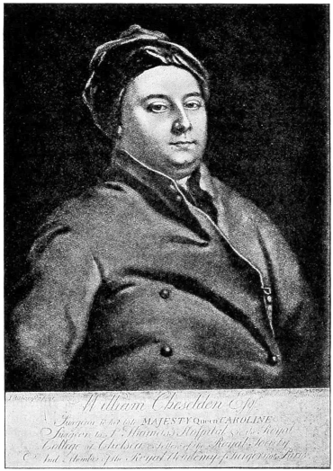
Portrait of William Cheselden, 1688–1752. Painted by Richardson.
The Project Gutenberg eBook of History of Iridotomy, by S. Lewis Ziegler
Title: History of Iridotomy
Knife-Needle vs. Scissors—Description of Author’s V-Shaped Method
Author: S. Lewis Ziegler
Release Date: January 7, 2022 [eBook #67117]
Language: English
Produced by: Thiers Halliwell, deaurider and the Online Distributed Proofreading Team at https://www.pgdp.net (This file was produced from images generously made available by The Internet Archive)
The text of this e-book has mostly been preserved in its original form, including some archaic spellings. A composite illustration on page 25 showing surgical knives lined up vertically side by side has been split into its individual components in order to display the instruments in horizontal orientation along with their respective captions. Hyperlinks have been added to textual cross-references and to footnotes. Page numbers are shown in the right margin and footnotes are located at the end. Footnotes are listed at the end.
The cover image of the book was created by the transcriber and is placed in the public domain.
3
HISTORY OF IRIDOTOMY.
KNIFE-NEEDLE VS. SCISSORS—DESCRIPTION OF AUTHOR’S
V-SHAPED METHOD.1
S. LEWIS ZIEGLER, A.M., M.D., Sc.D.
Attending Surgeon, Wills Eye Hospital; Ophthalmic Surgeon,
St. Joseph’s Hospital.
PHILADELPHIA.
To Cheselden has been conceded the honor of being the father and originator of iridotomy. Nearly two centuries have elapsed since he first published the report of his procedure in the Philosophical Transactions for 1728. Ever since that time, his signal success has been acknowledged by all except those who either failed to equal his dexterity, or who were prejudiced by their ambition to originate a new method.
A careful review of the medical literature of the century and a half following Cheselden’s announcement can not fail to impress the reader with the great interest attached to operations for the formation of an artificial pupil, which subject was considered second only in importance to that of cataract itself. Not only were a large number of monographs devoted wholly to this subject, but every work on general surgical topics set aside one or more chapters for the discussion of artificial pupil. This is in great contrast to the limited space which modern works on ophthalmology grudgingly yield to this still important subject.
It is difficult for us to appreciate the conditions which brought about so large a percentage of cases of pupillary occlusion. Crude surgical procedures, poor operative technic and the utter lack of asepsis often resulted in iridocyclitis or iridochorioiditis. The couching of the4 lens, the free discission of both hard and soft cataracts, the frequent introduction of the knife-needle through the dangerous ciliary zone, and the bungling efforts at extraction all increased the tendency to inflammatory reaction, while inadequate therapeutics and lack of antiphlogistic measures frequently permitted the deposit of plastic exudate in the pupillary area, thus resulting in membranous occlusion of the pupil.
For the sake of historical completeness, and in order to better emphasize the special domain of iridotomy, I will mention briefly the various methods that have been employed in making an artificial pupil. These are:
(1) Division of the thickened iris-membrane by an incision made either through the sclerotica or through the cornea. This is true iridotomy.
(2) Excision of a portion of the iris through a previously made corneal opening. This is now known as iridectomy.
(3) Separation of the iris from its ciliary attachment. This was generally known as iridodialysis, but sometimes called iridorrhexis.
(4) Simple incision of the pupillary margin, and of the free iris tissue. This has been designated sphincterotomy by some, and coretomy or iritomy by others. Either one of the latter terms is to be preferred, because it is more clearly descriptive.
(5) Detachment of the synechiæ at the pupillary margin, either anterior or posterior, thus allowing the pupil to retract. This was known as corelysis.
(6) Strangulation of the prolapsed iris in the corneal incision was called iridencleisis. The prolapse was sometimes tied with a ligature.
(7) Trephining of the iris-membrane, by passing a small trephine or punch through a corneal incision.
(8) Section and removal of a portion of the sclerotica and chorioid by knife or trephine, with replacement of the conjunctiva over this opening, the conjunctiva thus acting as a substitute for the cornea in transmitting light. This was called sclerectomy.
(9) Transplantation of the cornea for total leucoma. This was usually preceded by partial or complete trephining of this membrane.
5
In addition to these nine distinct methods certain combinations of these have been described and successfully practiced:
(10) Division and excision have frequently been performed together.
(11) Separation and excision have likewise had some vogue.
(12) Separation and strangulation have occasionally been practiced.
(13) Detachment of the synechiæ and excision have also been performed.
In this brief review of iridotomy,2 we shall confine our attention to the methods that have been advanced for the formation of an artificial pupil in cases of membranous occlusion of the pupil following removal of the lens, either by couching, extraction or discission, the iris-membrane in these cases being chiefly composed of inflamed iris tissue glued down by retro-iridian exudate to the thickened lens capsule.
The early history of iridotomy shows that the advocates of this operation were divided into two schools, (1) those recommending the use of the knife-needle for incising the iris-membrane, and (2) those adopting the method of introducing scissors through a previously made corneal section and freely incising the iris-membrane, or excising a portion of the same. We will first consider the school which advocated incision by the knife-needle.

Portrait of William Cheselden, 1688–1752. Painted by Richardson.
Cheselden,3 a renowned surgeon, and oculist to Her Majesty, Queen Caroline of England, first announced, in 1728, his success in making an artificial pupil by means of his knife-needle. He made his puncture back of the corneoscleral junction on the temporal side, passing the knife across the posterior chamber, and making a counter-puncture in the iris-membrane near the nasal margin. He then cut through the iris from behind forward as he withdrew the knife, the incision being carried through two-thirds of its extent.6 The pupillary opening thus made was a long oval slit, horizontally placed. He has reported two successful cases4 (Figs. 1 and 2), occurring in patients who had previously undergone couching of the lens. His instrument, strange to say, was practically of the same general shape as the Hays knife-needle, but was larger, and judging from the description more clumsily constructed, as there was danger of leakage of the aqueous and sometimes of the vitreous when it was used. Its form resembled a combination of a bistoury and a sickle-shaped7 knife, having a sharp edge on one side, a rounded back, and an acute point. We possess two good illustrations of this knife-needle, one by Cheselden himself (Fig. 3), and the other by his pupil, Sharpe5 (Fig. 4).
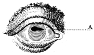
Fig. 1.—Original case of iridotomy. Iris incised above (Cheselden).

Fig. 2.—Second case of iridotomy. Iris incised below (Cheselden).

Fig. 3.—Original knife-needle in situ, behind the iris (Cheselden).
For more than a century the method of Cheselden seems to have been the storm center of controversy. Some doubted his veracity, others essayed his operation but failed, while a few had a moderate degree of success. Many attributed to him statements which do not appear in his published report. He says clearly that in each of his cases couching had previously been performed, and yet some have insisted that the lens was present, and must have been wounded. He also states that his incision was made from behind forward, and yet his followers, Sharpe5 and Adams,6 both describe the incision as being made from before backward. As Sharpe was his pupil, and presumably had seen him operate, Guthrie7 suggests the possibility of his having made his incision both ways, the technic being practically the same.
8
Morand,8 in his “Eulogy of Cheselden,” claims to have personally seen him operate “on an eye in which the iris was closed by an accident,” and gives a more detailed description which closely follows the original method. He states that Cheselden presented him with one of his knife-needles as a souvenir of the occasion. Although Morand does not record the exact date of his visit to London, he does state that it occurred during the year 1729. Huguier,9 in his exhaustive thesis on artificial pupil, also places the date of this visit in the year 1729. This fact is important, as some writers have declared that Morand neither made the visit to London nor saw Cheselden operate, but only quoted the original account given in the Philosophical Transactions. The publication of Morand’s high encomiums in 1757 attracted renewed interest to the subject of Cheselden’s operation among men of scientific and medical attainments.
Sharpe,5 in 1739, performed this operation in the same manner as Cheselden, except that after he had entered the knife-needle through the sclerotic he passed it through the iris and across the anterior chamber, and then incised the iris-membrane from before backward. Although he was Cheselden’s pupil, and dedicated his small volume on surgery to him, he probably did his master more harm than good, as all the objections to Cheselden’s method seemed to be based on the deprecatory remarks of Sharpe. He says, “I once performed it with tolerable success, and a few months after, the very orifice I had made contracted and brought on blindness again.” He mentions the danger of wounding the lens, the lack of success in paralytic iris with affection of the retina, the danger of iridodialysis from traction of the knife, and the possibility of failure because the incision would not enlarge sufficiently. Thirty years later (1769) he published the ninth edition of his book without recording a single additional case, but added the thought that, since extraction of the crystalline lens showed the cornea was not so vulnerable as had been believed, he would “imagine” that a larger knife might be introduced perpendicularly through the cornea and iris and a similar incision made. In his first eight editions he pictures Cheselden’s9 iris-knife (Fig. 4, vide p. 25), but in his ninth edition he substitutes a broad lance-knife with two edges which closely resembled the one Wenzel (vide Fig. 17) had just introduced (1767), and which Sharpe suggests “can also be used for the extraction of the cataract.” He evidently did not have a very clear idea of the subject, and only succeeded in casting doubt and discredit on the method of Cheselden, which, judging by his own statement, he had tried but once.
Heuermann,10 in 1756, had already antedated these thoughts of Sharpe by practising a similar method. He passed a double edged lance-knife through the cornea instead of through the sclera, and then made a sweeping incision through the iris-membrane without enlarging the corneal wound. He was probably the first to puncture the cornea with the iris-knife.
Janin,11 about 1766, performed Cheselden’s operation several times with but little success owing to reclosure of the wound by plastic exudate. He adopted Sharpe’s modification, but later on changed the incision from a horizontal to a vertical one with better results. He, however, afterward abandoned this procedure and became the originator of the other school, composed of those who preferred to use the scissors.
Guérin,12 in 1769, made a free corneal incision with a large cataract knife, and then introduced a small iris-knife, with which he made a crucial incision from before backward in the center of the iris-membrane. Although Guthrie7 distinctly states that Guérin afterwards removed the four angles of the cross with a pair of scissors in order to prevent reclosure of the incision, no direct confirmation of this statement can be found in his writings.
Beer,13 in 1792, first published his method, which he designated as “an improvement on Cheselden’s method.” Although the technic is somewhat different, the procedure is practically the same as that originated by Heuermann in 1756. Beer selected certain cases in which a prolapsed iris had followed the lower incision for cataract, causing adherent leucoma with a tensely10 drawn iris-membrane. He plunged his double-edged lance-knife (Fig. 5) through the cornea and stretched out iris, from above downward and a little obliquely (Fig. 6), so as to incise the center of the tense iris fibers crosswise, at right angles to the line of traction; cutting horizontally when the traction was vertical, and vertically when this was horizontal. In his monograph on artificial pupil,14 1805, he substitutes for the lance-knife his new broad iris-knife, which is practically the same as that later shown by Walton (vide Fig. 12), as, indeed, Walton’s procedure (vide Fig. 13) was almost identical with that of Beer. For other conditions he usually employed Wenzel’s operation until by chance he encountered a puzzling case which led him to perform the operation we now know as iridectomy (1797) and which thereafter became his favorite procedure for artificial pupil.
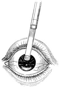
Fig. 6.—Beer’s iridotomy with broad iris-knife (after Mackenzie).
Adams,15 in 1812, revived the operation of Cheselden with certain modifications. While his puncture was made in the same location, his technic was different. He entered the sclera with a small iris-scalpel of his own special design (Fig. 7), which, like Sharpe, he passed through the iris-membrane into the anterior chamber,11 carrying it across to the nasal side (Fig. 8). From entrance to exit he always kept the edge of the knife turned back toward the iris, so as to cut from before backward. He was thus able by the most delicate pressure of his instrument, to make a long horizontal incision, without causing iridodialysis (Fig. 9). If the first incision appeared to be too short, he did not withdraw the knife entirely, but again carried it forward and partially withdrew it, always cutting in the same plane. To quote his own words, “by repeating the efforts to divide the iris (taking care in so doing to make as slight a degree of pressure as possible upon the instrument, instead of withdrawing it out of the eye at once, as recommended by Cheselden), a division of that membrane may, in almost all cases be effected, of a requisite size to establish a permanent artificial pupil” (Figs. 10 and 11).

Fig. 8.—Adams’ iris scalpel in situ, showing location of scleral puncture (after Lawrence).

Fig. 9.—Iridotomy by Adams’ method (after Lawrence).
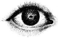
Fig. 10.—Occlusion of pupil (Adams).
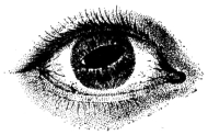
Fig. 11.—The resulting pupil after iridotomy (Adams).
Here were three elements of success, a sharp knife, a gentle sawing movement, and the most delicate pressure of the instrument. His method was a decided advance, and he reported success in nearly one hundred cases. Others, less skilful, however, failed of success, and the severe criticisms of Scarpa,16 though evidently unjust and tinged by personal animosity,17 cast a shadow of doubt on the method.
12
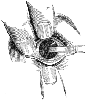
Fig. 13.—Iris-knife in position to make central pupil (Walton, after Beer).
From that time on for nearly half a century this form of iridotomy was practically abandoned, the pendulum swinging toward the use of scissors, which Maunoir had popularized and Scarpa had indorsed. Walton,18 however, about 1852, proposed a method closely resembling that of Heuermann and almost identical with that of Beer (vide Fig. 6). His iris-knife (Fig. 12) was practically the same as the broad iris-knife of Beer. He incised the cornea near the limbus, and passed the knife across the anterior chamber to the middle of the iris-membrane which he punctured with a sweeping vertical incision (Fig. 13). If the tissue still retained its elasticity there appeared a long pupillary aperture, elliptical and vertical (Figs. 14 and 15). This incision, however, like all those made through a single set of the iris fibers, was only successful when there was sufficient resiliency remaining in the iris tissue to draw the slit open, and thus keep the edges from uniting. While this method never became very popular, there were some who later practiced it by substituting a very narrow Graefe knife for the iris-knife of Heuermann, Beer and Walton. In fact, this latter procedure still has considerable vogue, both for iridotomy and capsulotomy.
13
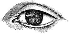
Fig. 14.—Occlusion of pupil (Walton).
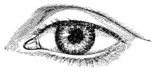
Fig. 15. New pupil after incision with iris-knife (Walton).
During the following seventeen years no notable advance was made, the scissors method still retaining its hold on the profession, until in 1869, von Graefe, after long reflection, became convinced of the dangers of that method, and communicated to one of his pupils, M. Meyer, his method of simple iridotomy performed with the knife-needle. Meyer19 quotes his views as follows:
“For such cases von Graefe has suggested another method of operation, the principle and execution of which are contained in the following note written for us by that illustrious savant in 1869:
“When, in consequence of a cataract operation, the lens is absent, and when there is highly developed retro-iritic exudation, with disorganization of the iris tissue, flattening of the cornea and the other sequelæ of a destructive iridocyclitis, I substitute simple iridotomy for iridectomy, which is the operation hitherto performed, generally without success. The operation consists in inserting a double-edged knife, resembling in shape a very sharp pointed lance-knife, through the cornea and newly formed tissues till it pierces the vitreous body, and immediately withdrawing it; and, while withdrawing it, enlarging the wound in the membranes without increasing the size of the corneal wound. Experience shows that such plastic membranes attached to the atrophied iris and to the capsule of the lens have a tendency to contract sufficient to maintain, to a certain extent, the opening which has been made.
“If, in the ordinary method of iridectomy, combined with laceration or extraction of the false membranes, we find that the artificial pupil usually becomes closed, we must attribute this to an excessive vulnerability, which immediately sets up proliferation in those tissues which have been touched, and which are endowed, in consequence on their structure, with an irritability altogether peculiar. We know that even the transitory reduction of the intraocular pressure, which follows the evacuation of the aqueous humor, is sufficient to give rise to14 hemorrhage in the anterior chamber, which interferes with the perfect success of the intended operation; but most of our failures in the ordinary methods are due to the irritation caused by the forceps and the traction on the surrounding structures. Simple iridotomy is free from such inconveniences; it is, so to speak, a sub-corneal act, and enjoys the immunity which belongs to subcutaneous operations.
“I have also reduced the corneal wound to a minimum, by using small falciform knives. These are passed through the false membranes, which are then cut from behind forward.”
Von Graefe thus proposed two methods, (1) by cutting from before backward with a double-edged lance-knife, according to the method of Heuermann, and (2) by cutting from behind forward with a sickle-shaped knife, after the original suggestion of Cheselden. Later in the same year, as he lay on his last bed of illness, he became so absorbed in the study of this subject that he sent a telegram to the Heidelberg Congress20 (September, 1869), in which he advocated the method by the sickle-shaped knife-needle as the best procedure. His last message to his colleagues showed, therefore, that through mature conviction he strongly favored the use of the knife-needle, and the making of a sub-corneal incision in the iris-membrane without evacuating the aqueous humor. His untimely death, however, prevented him from further perfecting this procedure and presenting it to the profession.
Galezowski,21 in 1875, published a somewhat similar method in which he used his falciform knife, aiguille-a-serpette (Fig. 16), which he introduced through the cornea and iris-membrane, making either a horizontal or a vertical incision, with a “go-and-come” (sawing) movement, after the suggestion of Adams. If this single cut was not sufficient, he made a linear incision of the cornea with a Graefe knife, drew out the iris and cut it off with scissors. By a process of evolution, however, he perfected the former procedure and eliminated the scissors. This latter method was published in the third edition of his book in 1888. He punctured the cornea and iris-membrane with the sickle-shaped knife, making first a horizontal incision by the sawing movement of Adams, and finishing with a second cut in the vertical direction, thus forming a T-shaped incision. In actual15 practice, however, he almost always prolonged this second cut, thus making a crucial incision after the manner of Guérin.12
The writer,22 in 1888, was led to devise an operation with a modified Hays knife-needle, in which through a corneal puncture he made a converging incision in the iris-membrane which resembled an inverted V. The resulting pupil opened up and formed either a triangular or an oval-shaped pupil depending on the degree of stiffness or resiliency of the iris-membrane. This method will be described in detail later on.
We will now return to the consideration of the second school in which scissors were introduced through a previously made corneal section and a free incision was made in the iris-membrane, or a portion of the membrane excised.
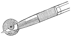
Fig. 17.—Wenzel’s cataract knife, and method of incision (after Mackenzie).
Janin,11 in 1768, having abandoned the procedure of Cheselden, proposed a new method. He incised the cornea below as for cataract extraction, and raised the corneal lip with a spatula while he introduced a pair of curved scissors, the lower blade of which was pointed. He plunged this sharp blade through the iris-membrane, and with a single vertical cut made a crescentic pupil which gaped sufficiently for visual purposes. As this is the first known description of iridotomy by the scissors method it is probable that Janin was the originator of this procedure.
Wenzel,23 in 1786, employed a different method. With a lance-shaped cataract knife he entered the cornea,16 dipped through the iris-membrane, returned to the anterior chamber, and continuing to cut made a counter-puncture on the opposite side of the cornea, following which he completed his cataract incision. This gave a semilunar flap of iris tissue which could easily be excised by scissors passed through the large corneal opening (Fig. 17).
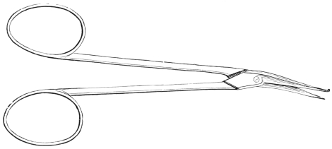
Fig. 18.—Maunoir’s scissors.
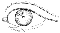
Fig. 19.—V-shaped iridotomy with scissors (Maunoir).

Fig. 20.—Parallelogram pupil (Maunoir).
Maunoir,24 in 1802, took up the method of Janin, with the object of improving it. He made an incision near the corneal margin, through which he introduced a pair of long, thin, angular scissors of his own design (Fig. 18), one blade of which was sharp-pointed like a lancet, and the other button-pointed like a probe. The iris-membrane was then punctured by the sharp blade at about the natural location of the pupil, and an incision executed toward the ciliary margin of the iris. Finding that this single incision did not always succeed,25 he subsequently improved this method by making a second incision from the pupillary area toward the iris margin, in the line of the radiating iris fibers, thus making a divergent V (Fig. 19). This triangular flap was then allowed to shrink back, or if too stiff, was17 drawn out and excised. The resultant pupil assumed the shape either of a triangle, a parallelogram (Fig. 20), or a crescent (Fig. 21). He always made his incision parallel with the radiating fibers of the iris and across the circular fibers.
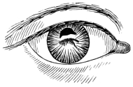
Fig. 21.—Crescent pupil (Maunoir).
Scarpa,16 in 1818, having abandoned his own method of iridodialysis as wholly unsatisfactory, adopted Maunoir’s procedure with enthusiasm, chiefly because he had by a friendly correspondence25 personally encouraged Maunoir with advice and suggestion during its development. He indorsed Maunoir’s plan of a double incision when he stated his conviction that “experience has proved that in order to obtain, with the most absolute certainty, a permanent artificial pupil, it is necessary to make two incisions in the iris so as to form a triangular flap in the membrane, in the form of a letter V, the apex being precisely in the center of the iris and the base near the great margin.” Some have claimed that Scarpa himself originated the V-shaped incision, but he gives Maunoir full credit for its successful accomplishment, although he does suggest some additional indications for its practical application.
His opposition to the knife-needle incision of Cheselden arose from the fact that the pupil either did not open, or if it did open would not remain permanent, chiefly because of the single iris incision. His antagonism to the more successful procedure of Adams was the result of a caustic personal controversy17 with that skilful surgeon, who ably parried his charges.15 His great influence with the profession of that day, however, served to check the sentiment in favor of Adams’ procedure, and when the weight of his indorsement was cast in favor of Maunoir’s operation the scales were decisively turned toward the side of the scissors method.
18
Mackenzie,26 in 1840, practiced Maunoir’s operation with considerable success, but in certain cases found it necessary to employ a slight modification of this procedure. He reversed Maunoir’s incision by making the same divergent V across the radiating fibers of the iris instead of parallel with them (Fig. 22), thus securing a triangular pupil (Fig. 23), which Lawrence27 thought might succeed in some cases where Maunoir’s method would not be available.

Fig. 22.—Mackenzie’s incision in cornea and iris-membrane (Mackenzie).

Fig. 23.—Resulting triangular pupil from Mackenzie’s incision (Mackenzie).
Bowman,28 in 1872, proposed a method which, though surgically difficult to execute, was quite ingenious, and may have been the initial suggestion that stimulated DeWecker to write his monograph in the following year. I will quote his description as follows: “We make a double opening simultaneously on opposite sides of the cornea. It is more convenient, of course, to make these two openings in a horizontal than in a vertical direction. I then run a pair of scissors in two diverging lines (V) from each incision, thus enclosing between the incisions a large square or rhomboidal portion of the iridial region including the pupil, and all the structures there. You then withdraw the portion thus cut out. There is no drag on the ciliary region; whatever is withdrawn has been cut away from its connections beforehand” (Figs. 24, 25 and 26).

Fig 24.—Plan of Bowman’s first iris incision. Divergent V.

Fig. 25.—First incision completed. Plan of second, showing double V.

Fig. 26.—Rhomboidal pupil, resulting from Bowman’s iridotomy.
19
This method is simply an elaboration of the one proposed by Maunoir, in which, instead of forming one divergent V, Bowman has made a duplicate incision on the opposite side, and by joining the bases of these two resultant triangles has caused them to take the shape of a rhomboid, thus <>.
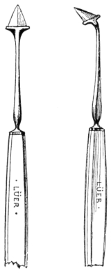
Fig. 27.—Stop keratomes, straight and angular (De Wecker).
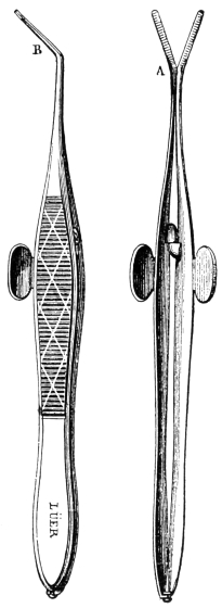
Fig. 28.—Forceps-scissors (pinces-ciseaux) (DeWecker).
DeWecker,29 in 1873, published his admirable monograph on iridotomy, in which he proposed the operation which bears his name, and which has long stood as the best recognized method of this procedure. He advocated20 two different ways of performing this: 1, simple iridotomy, and 2, double iridotomy.
1. Simple Iridotomy.—This is practically the same operation as Critchett’s sphincterotomy and Bowman’s visual iridotomy, although differently executed. It has been supplanted in our day by iridectomy, and does not, therefore, come within the purview of this discussion.
2. Double Iridotomy.—He rightly claimed that this was both antiphlogistic and optical in its purpose. He employed two distinct methods, which he designated as (a) iritoectomie, and (b) iridodialysis. The instruments he used were a small stop-keratome (Fig. 27) and a pair of specially devised fine iris scissors (pinces-ciseaux) (Fig. 28), one blade being sharp pointed and the other blunt. These scissors were a great mechanical advance over all previous instruments of this kind, and undoubtedly proved to be a most important element in the success of his procedure.

Fig. 29.—Iritoectomie. Convergent V (DeWecker).

Fig. 30.—Iridodialysis. Divergent V (DeWecker).
(a) Iritoectomie.—He entered the stop-keratome through the cornea, made an exact 4 millimeter incision, and then partly withdrew it while letting the aqueous slowly escape. As soon as the iris-membrane floated up against the knife, he pressed forward, making a 2 millimeter incision in the iris. Slowly withdrawing the knife, he introduced the sharp point of the scissors through the iris buttonhole and cut obliquely from either extremity of the incision toward the apex of a triangle, thus making a convergent V (Fig. 29). He then grasped the resulting triangular flap with the forceps and removed it, leaving an open central pupil.
(b) Iridodialysis.—His second method was a counterpart of Maunoir’s earlier operation, with the addition of iridodialysis. He made the corneal and iris incision with the stop-knife, as in the previous method. Slipping in his scissors he cut from the center of the iris-membrane toward the periphery, and duplicated this incision at an oblique angle to the first, thus making a divergent V (Fig. 30). This formed a triangular flap21 which he grasped with forceps and tore from its ciliary attachment by iridodialysis.
DeWecker’s procedure was planned by a skilled operator, and required great dexterity in its execution. When successful, however, the result was most brilliant. Nevertheless, it was impossible to eliminate the danger of hemorrhage and loss of fluid vitreous in iritoectomie, while in iridodialysis there was the added danger of a torn ciliary surface and traction on the ciliary body. His strict injunction to have a trained assistant hold up the speculum blades in order to avoid the loss of fluid vitreous, showed how much he feared this disastrous contretemps. The success of his method of incision is well shown in the illustration of his two cases (Figs. 31 and 32).
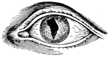
Fig. 31.—Pupil by iritoectomie. Two incisions. Convergent V (DeWecker).
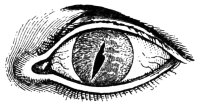
Fig. 32.—Stenopaic pupil. Single iris incision (DeWecker).
I have already suggested the possibility of Bowman’s paper before the London Congress of 1872 having given origin to DeWecker’s monograph in 1873. This seems quite reasonable when we consider that Bowman proposed two methods of iridotomy, one his double V operation with a rhomboidal pupil (previously quoted), and the other a visual iridotomy or sphincterotomy, by cutting through the pupillary margin with a blunt corneal knife. These two methods are exact prototypes of DeWecker’s proposals. Furthermore, DeWecker was present at the London Congress where he heard Bowman’s paper, and took part in its discussion. In fact, thirteen years later DeWecker acknowledged30 that after considering the objections to Bowman’s method of iridotomy “I addressed myself at that time to the search for an instrument which allows the avoidance of all traction on the iris, and which can be handled through a narrow opening, while exerting its cutting action in a22 plane parallel to the surface of the cornea, against which the diaphragm of the iris applies itself, after the escape of the aqueous humor. The forceps-scissors having been discovered, it was easy for me to cause to be revived the procedure of Janin, and to make it decisively take rank in modern ocular surgery.”
DeWecker makes only a casual reference to Maunoir’s method, but credits Janin with the original suggestion of the method which he has thus elaborated. Nevertheless, it is quite evident that DeWecker’s method was simply a modification of the one outlined by Maunoir seventy years before. Furthermore, he lays down the same rule that Maunoir first offered: “Always cut parallel to the radiating fibers and perpendicularly to the circular fibers of the iris.”
In reviewing the questions at issue between these two schools of iridotomy, one can not help noticing the constant oscillation from one method to the other as certain advances were made. The method by the knife-needle seemed to possess the advantage of easy accomplishment and less postoperative disturbance, but with the disadvantage that often the pupillary opening was inadequate and promptly reclosed by plastic exudate. On the other hand, the method by the scissors was more difficult of accomplishment, caused more traumatism to the eye, was often complicated by great loss of fluid vitreous, and was frequently followed by severe inflammatory reaction. If, however, it proved successful, the resulting pupil was permanent and sufficiently large for visual purposes. The inclination of all operators seemed to be toward the use of the knife-needle, and it was only necessity that forced them to adopt the more complicated procedure of the open operation with scissors. Von Graefe seemed to recognize this when he referred to the knife-needle incision as “a sub-corneal act which enjoys the immunity of subcutaneous operations.”
The chief advantages of iridotomy by the knife-needle are the ease of incision, the lack of traction on the ciliary body, the freedom from postoperative inflammatory reaction, the avoidance of opening an eyeball which may contain fluid vitreous, the lessening of the tendency to iris hemorrhage from lowered tension, and the avoidance of the nebulous scar which often follows23 a large corneal incision in old inflammatory eyes. The disadvantages revealed in the method of the knife-needle lay partly in the method and partly in the faulty instruments constructed in that day. Cheselden, Morand, Sharpe and Adams all made the mistake of entering the eye back of the corneoscleral junction, which is so near to the danger zone of the eye. Adams, however, made a two-fold improvement in adding to his operation a sawing movement and in advocating the “most delicate pressure of the instrument” in order to make a free incision. Heuermann was apparently the first to make the puncture through the cornea instead of through the sclera.
The advocates of the knife-needle method long labored under the disadvantage of making a single iris incision, while those who employed the scissors early discovered that a double incision was necessary to success. Although Janin was the originator of the scissors method, Maunoir was the first to deliberately try a triangular flap, which DeWecker later elaborated and made a permanent success. The many disastrous results of the open operation, however, compelled conservative surgeons, like von Graefe, to revert to a study of Cheselden’s method, and to seriously consider the great advantages which a successful iridotomy by the knife-needle method would confer on surgeon and patient alike.
1. Cheselden’s knife-needle (Figs. 3 and 4) was a splendidly designed instrument, but a poorly executed one. The blade was too large (11 mm.) and the shank improperly rounded, so that both aqueous and vitreous were liable to escape through the scleral puncture. This leakage may explain many failures, although the single iris incision was undoubtedly the most serious fault of the method.
2. The iris-scalpel of Adams (Fig. 7) was poorly designed but splendidly executed, the long blade completely filling the wound and thus preventing the escape of any fluid. The cutting edge, however, was too long (15 to 20 mm.), and especially so for the execution of the sawing movement advised by Adams.
3. The double-edged lance-knife (Figs. 5, 12 and 33) employed by Heuermann, Beer and von Graefe, was useful for the long sweeping incision in the iris-membrane24 which they advocated, but is not adapted for the method which will be described later. The same shaped knife (Fig. 33) with a smaller blade and a longer shank is also used for this purpose, but is likewise too broad, too oval pointed and too much bellied to cut well, while the upper edge is liable to scarify Descemet’s membrane at the same time that the lower edge is executing the incision in the iris tissue.
4. The sickle-shaped knife (Fig. 16) which von Graefe recommends and Galezowski employs, is excellent for making the puncture, but for the go-and-come movement, which Galezowski advises, is not nearly so good as the straight blade with a slight falciform point. It closely resembles the older falciform knife of Scarpa.
5. The knife-needle of Knapp (Fig. 34), which is so generally used for capsulotomy, is unfortunately not well adapted for iridotomy. The point is too oval, the cutting edge is too much bellied, and the blade is too short (5 mm.). It will not easily puncture a dense iris-membrane, and the long sawing incision can not be well executed, because the short blade either persists in slipping out of the iris incision or else allows the membrane to ride up on the shank, in either case interfering with the completion of the operation.
6. Sichel’s iridotome (Fig. 35) closely resembles Knapp’s knife-needle, and although specially designed for this purpose, has the same faults, an oval point and a bellied edge. On the other hand, the blade is too long (11 mm.) to be easily manipulated in the anterior chamber.
7. The Hays knife-needle (Fig. 36), as suggested in the early part of this paper, has the same general shape as Cheselden’s instrument, although much smaller. It was devised by Dr. Isaac Hays, an early surgeon of the Wills Hospital, and, although not well known to the profession at large, has been in constant use by the staff of that hospital for more than half a century. I may be pardoned for briefly quoting the original description of the instrument as published by Hays31 in 1855:
25
“This instrument from the point to the head, near the handle (a to b, Fig. 36), is six-tenths of an inch, its cutting edge (a to c) is nearly four-tenths of an inch. The back is straight to near the point, where it is truncated so as to make the26 point stronger, but at the same time leaving it very acute, and the edge of this truncated portion of the back is made to cut. The remainder of the back is simply rounded off. The cutting edge is perfectly straight and is made to cut up to the part where the instrument becomes round, c. This portion requires to be carefully constructed, so that as the instrument enters the eye it shall fill up the incision, and thus prevent the escape of the aqueous humor.”

Fig. 4.—Cheselden’s knife-needle (after Sharpe).

Fig. 37.—Ziegler’s model of knife-needle.

Fig. 36.—Hays’ knife-needle, exact size and enlarged (Hays).

Fig. 16.—Sickle-shaped knife, Aiguille-à-serpette (Galezowski).

Fig. 35.—Sichel’s iridotome (after Meyer).

Fig. 34.—Knapp’s knife-needle.

Fig. 7.—Adams’ iris-scalpel; large and small size.

Fig. 33.—Double edged lance-knife (modern model).

Fig. 5.—Double edged lance-knife (Beer).

Fig. 12.—Iris-knife (Walton, after Beer).
The Various Knife-Needles and Iris-Knives Mentioned in the Text.
(Grouped together for study and comparison.)
8. The knife-needle, which I invariably use, is a modified pattern of that devised by Hays. The form of this instrument lies midway between the falciform knife and the bistoury, and possesses the advantages of both. It has a very delicate point which punctures easily, and an excellent cutting edge of sufficient length (7 mm.). If the shank is properly rounded it can be used with a sawing motion, sliding backward and forward through the corneal puncture without injuring the cornea, and without allowing the aqueous to escape. To accomplish this the more easily, the shank has been made 4 mm. longer than the original model. This instrument, therefore, seems to meet all the requirements of a perfect iris-knife, viz., a falciform point which makes the best puncture, a straight edged blade which makes the best incision, and a cutting edge 7 mm. long, which is the best length for properly executing the sawing movement. My model32 of knife-needle (Fig. 37) resembles Cheselden’s knife, as shown by Sharpe (Fig. 4), even more closely than the original pattern of Hays does.

Fig. 37.—Ziegler’s model of knife-needle.
1. A good knife-needle must be carefully selected. We have already concluded that the modified Hays knife-needle is the best model for this purpose. The knife-needle must, of course, have a well sharpened point and edge.
2. The character of the incision in the iris-membrane is of vital importance. It should be a double incision. Guérin, Maunoir, DeWecker and Galezowski recognized27 this. Guérin made a crucial incision, Maunoir and DeWecker adopted the triangular flap, while Galezowski advocated the T-shaped cut. Our choice is the V-shaped incision, which is undoubtedly the only one that will cut through all the iritic fibers in such a way as to give us the greatest retraction of the membrane.
3. Absolutely no pressure should be made in cutting with the knife-needle. This must be recognized as the main secret of success, whether you are incising a dense, felt-like iris-membrane, or a thin filmy capsule. If this rule is observed all traction on the ciliary body will be avoided.
4. The knife-needle should slide backward and forward through the corneal puncture with a gentle sawing movement.
5. The corneal puncture and membrane counter-puncture should be far enough apart to make the corneal puncture a good fulcrum for the delicate leverage necessary in executing the iris incision.
6. The knife-needle should be so manipulated that no aqueous shall be lost, as this accident may prevent the completion of the operation, and may increase the tendency to iris hemorrhage by lowering the ocular tension.
7. Every incision should be made a thoroughly clean cut, and all tearing of the tissues should be avoided.
8. The most perfect artificial illumination should be secured, either by an electric photophore or a condensing lens, as both iridotomy and capsulotomy require constant and close inspection of the operative field.
The method of V-shaped iridotomy, performed by me with my modified Hays knife-needle, may be described as follows:
First Stage.—With the blade turned on the flat, the knife-needle is entered at the corneo-scleral junction, or through the upper part of the cornea (Fig. 38), and passed completely across the anterior chamber to within 3 millimeters33 of the apparent iris periphery. The knife is then turned edge downward, and carried 3 millimeters to the left of the vertical plane (Fig. 39).
Second Stage.—The point is now allowed to rest on the iris-membrane, and with a dart-like thrust the membrane28 is pierced. Then without making pressure on the tissue to be cut, the knife is drawn gently up and down with a saw-like motion, until the incision has been carried through the iris tissue from the point of the membrane puncture to just beneath the point of the corneal puncture. This movement is made wholly in a line with the axis of the knife, the shank passing to and fro through the corneal puncture, and the loss of any aqueous being carefully avoided in the manipulation.
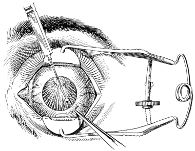
Fig. 38.—Author’s V-shaped iridotomy. Knife-needle entered through cornea.

Fig. 39.—Author’s method. Plan of first incision.

Fig. 40.—First incision completed. Plan of second incision.

Fig. 41.—Pupil resulting from V-shaped iridotomy.
Third Stage.—The pressure of the vitreous will now cause the edges of the incision to immediately bulge open into a long oval (Fig. 40) through which the knife-blade is raised upward, until above the iris-membrane, and then swung across the anterior chamber to a corresponding point on the right of the vertical plane, which, owing to the disturbance in the relation of the29 parts made by the first cut, is now somewhat displaced and the second puncture must be made at least 1 millimeter farther over, i. e., 4 millimeters to the right of the vertical plane (Fig. 40).
Fourth Stage.—With the knife point again resting on the membrane, a second puncture is made by the same quick thrust, and the incision rapidly carried forward by the sawing movement to meet the extremity of the first incision, at the apex of the triangle, thus making a converging V-shaped cut (Fig. 41). Care must be taken at this point that the pressure of the knife-edge on the tissue shall be most gentle, and that the second incision shall terminate a trifle inside the extremity of the first, in order that the last fiber may be severed and thus allow the apex of the flap to fall down behind the lower part of the iris-membrane. If the flap does not roll back of its own accord it may be pushed downward with the point of the knife. When the operation is completed the knife is again turned on the flat and quickly withdrawn.
The most fruitful sources of failure are, first, a poorly sharpened knife-needle; second, a badly planned incision; third, inability to sever the apex of the triangle; fourth, the early loss of aqueous; fifth, too heavy pressure with the knife-edge, and sixth, rocking or rotating the knife backward instead of making the sawing movement. All of these can easily be avoided, if the surgeon will only exercise care and good judgment.
In an occasional case, the iris-membrane may be so stiff that the apex of the flap will not retract. If the apex can not be pushed down by the tip of the knife turn the blade on the flat, puncture the base of the flap by a quick thrust, and with a sawing motion cut across its fibers so that it will fall back as though hinged; or, if positive that the vitreous is not fluid, introduce a keratome in the cornea below, draw out the triangular tongue, cut it off with the iris scissors, and dress back the base with a silver spatula.
It is possible that the capsule, or iris tissue, may lose its anchorage. In that event we must either reverse the procedure by entering the knife-needle below, and cut from above downward, or else pass a second knife-needle through the loosened edge of the membrane to fix it, and then proceed with the usual method.
30
Occasionally, the apex of the triangular flap will hold fast, because the last fiber of tissue has not been severed. If the leverage is too short to incise it from above, withdraw the knife-needle and reintroduce it far enough from the apex to secure the proper leverage, and again incise it gently, until it falls back.
Traction on the ciliary processes, accidental puncture of the ciliary body, or the tearing of the membrane from its ciliary attachment may all set up iridocyclitis or glaucoma, and should therefore be avoided. As tense capsular bands are liable to engender a similar condition they should be incised. If any of these traction bands should remain in the edge of the coloboma, we may enter the knife behind them and gently saw through into the already cleared pupil, before withdrawing the knife.
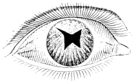
Fig. 42, (Case 1).—Iridotomy in a stiff iris-membrane (author’s original case).
I will briefly cite a few examples of the V-shaped operation, two that were my first efforts, and two that were recent cases. They were all of the class that are often abandoned as hopeless; hence the visual result is far below the operative success.
Case 1.—History.—F. M., aged 65 years. O. D. complete membranous occlusion of pupil from iridocyclitis, following cataract extraction. The iris and capsule are tensely drawn up toward the ciliary border. Light perception and projection good. Several efforts have been made to incise the membrane, but without success. Admitted to Wills Hospital by the late Dr. Goodman, through whose courtesy I operated.
Operation.—On Jan. 15, 1889, I made two long incisions, almost crucial, and extending beyond the apex of the V, resulting in a W-shaped pupil, on account of the stiff iris membrane (Fig. 42). With S. + 10 D. he saw 20/50.
Case 2.—History.—J. S., aged 30 years. O. S. injured and enucleated. O. D. sympathetic inflammation, chorioidal cataract; three discissions and one iridectomy, down and in. Membranous occlusion of pupil. I first saw him in 1888 while31 house surgeon at the Wills Hospital, where iridotomy was skilfully performed nine times by one of the surgeons, the methods being varied and ingenious, but without success, as the incision was invariably closed by plastic exudate. My interest in this series of operations first drew my attention to the subject of iridotomy, and stimulated me to develop the method I have here submitted and which I first tried in Case 1.
One year later this patient came to my clinic at St. Joseph’s Hospital. Iris was discolored, capsule thickened and visible through the coloboma, down and in; areas of scleral thinning, with pigmented chorioid showing through. T—3. Light perception good, projection only fair.
Operation.—On June 17, 1889, I made a V-shaped iridotomy along the outlines of the former iridectomy. The membrane freely opened up into a triangular or pear-shaped pupil (Fig. 43), which proved permanent, but was only useful for quantitative vision, about 5/200. No further test could be made because the disorganized vitreous was filled with floating masses. I have seen him within a year, going about and earning his living. From an operative standpoint I have always considered this early effort one of my most successful cases, chiefly because of the great density of the iris-membrane and the lowered tension of the eyeball.
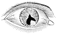
Fig. 43, (Case 2).—Iridotomy in a soft eyeball, with dense iris-membrane.
Case 3.—History.—Mrs. A. D., aged 45 years. O. D. iridectomy for glaucoma seven years ago. O. S. iridectomy two years ago by another surgeon, at which time there occurred slight incarceration of iris, followed by sympathetic ophthalmitis in O. D. The severe iridochorioiditis resulted in cataract and some shrinkage of globe. The cataracts were extracted from both eyes in 1907, followed by dense opacity of cornea above, iris bombé, shallow anterior chamber, T—2. Here was a soft, distensible, iris tissue with shallow anterior chamber and greatly lowered tension of the eyeball, constituting one of the most difficult conditions to operate on.
Operation.—On May 13, 1907, the eyes being quiet, and light perception and projection fair, V-shaped iridotomy was performed on both eyes. The leucomatous areas in the upper part of cornea necessitated making the pupil below. In O. D. the pupil opened up beautifully (Fig. 44), but in O. S. a tag32 of iris hung fast (Fig. 45) and was again incised two months later. The artist has illustrated the remaining portion of this tag very well. As soon as the iris tissue was incised it retracted, making the pupils larger than the area of incision. The test for glasses, nearly a year later, March 15, 1908, yielded the following result:
O. D. S + 13 D ⁐ C + 4.75 D ax. 105° = 20/40.
O. D. S + 13 D ⁐ C + 3 D ax. 65° = 20/40.
Add
O. D. S + 4 D = J. 10.
O. S. S + 4 D = J. 10.
These were ordered in biconvex torics. She had worn glasses for a year, but claims vision is much better with the new ones. This seems like an excellent result when we consider that these eyes had passed through glaucoma, iridochorioiditis and cataract, followed by membranous occlusion of pupil, lowered tension and fluid vitreous. The high hyperopia and astigmatism show the phthisical condition of each globe. There is marked cupping of both nerve heads and the fields are contracted.
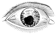
Fig. 44, (Case 3).—Iridotomy in a soft eyeball, with thin membrane and iris bombé.
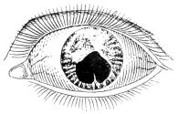
Fig. 45, (Case 3).—Iridotomy showing apex of iris flap after incision through adherent fibers.
Case 4.—History.—Mrs. B. M., aged 64 years. O. S. struck by a stone in childhood, destroying vision. Dense leucoma above, chorioidal cataract, calcareous deposit; exclusion of pupil. T—1. Lpc. good. Lpj. fair. O. D. recurrent attacks of inflammation for seven years, posterior synechiæ and cataract. Counts fingers at 6 inches. Extraction with iridectomy, both eyes, in 1907. Site of incision has become densely leucomatous. O. D. shows capsular area above, iris drawn up. O. S. complete membranous occlusion of pupil.
Operation.—Oct. 7, 1907, V-shaped incision was executed entirely in the iris tissue of O. D., the pupil spreading out into an ovoid shape (Fig. 46), leaving area of capsule and small band of iris above. O. S. was operated on Jan, 13, 1908, by the same method, the resulting pupil being almost round (Fig. 47) owing to the resilient iris tissue.
The test for glasses, March 10, 1908, gave the following result:
33
O. D. S + 12 D ⁐ C + 1.25 D ax. 135° = 20/50.
O. S. S + 12 D ⁐ C + 1.25 D ax. 135° = 20/70.
Add
O. D. S + 5 D = J. 6.
O. S. S + 5 D = J. 12.
These were ordered in biconvex torics, which she now wears with great comfort. It is worth noting that O. S. still retained good visual acuity, although blinded by an injury nearly fifty years before.
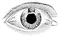
Fig. 46, (Case 4).—Irido-capsulotomy, with band of iris, and capsule in coloboma above.
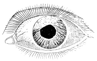
Fig. 47, (Case 4).—Iridotomy with round central pupil in a resilient iris-membrane.
The application of the V-shaped method to capsulotomy shows an even greater field of usefulness, as this method is par excellence the best way of incising a delicate secondary capsular cataract. This should be done under artificial illumination. The pupil should be dilated, as the area of incision is necessarily smaller than in iridotomy, and unnecessary wounding of the iris should be avoided. The proposed capsular opening must be so calculated as to fall within the area of the undilated pupil, or partly within the coloboma if an iridectomy has been previously performed.

Fig. 48.—Author’s Vshaped capsulotomy. Plan of first incision.

Fig. 49.—First incision completed. Plan of second incision.

Fig 50.—Pupil resulting from V-shaped capsulotomy.
The knife-needle is entered at the upper corneal margin, passed across the anterior chamber to a point 2 mm. to the left of the vertical plane (Fig. 48), the capsule34 punctured by a quick thrust, and the saw-like incision carried from below upward, as in iridotomy. The knife is then raised up above the capsule and swung 3 mm. to the right of the vertical plane (Fig. 49), the capsule is again punctured, and a duplicate incision carried up to join the first, at the apex of the converging V (Fig. 50).
Where the pupillary margin is adherent to the underlying capsule, or the pupillary space is too small, it may be necessary to start the incision in the iris tissue, a little below the pupil, and then cut upward until the knife emerges into the pupillary area, thus making an irido-capsulotomy. The soft iris tissue is easily incised if no pressure is made with the knife, and the sawing motion is maintained.
Postoperative inflammatory reaction is infrequent, but if it should occur the usual antiphlogistic treatment of atropin, calomel, ice-pads and leeching should be actively instituted and continued until the eye is absolutely quiet. The operation itself is frequently an antiphlogistic measure, because it relieves iris-tension and traction on the ciliary body. The usual compress of gauze and cotton, covered with a Liebreich patch, may be applied to the eye for the first twenty-four hours and rest in bed enjoined for that period.
We have carefully reviewed the history of iridotomy for nearly two centuries, and noted how the pendulum has swung from knife-needle to scissors, and back again. We have learned that Cheselden, the father of iridotomy, originated the method of incision by the knife-needle, which Heuermann modified, and Adams later revived and improved. We have seen how Janin abandoned this procedure and originated the scissors method, which Maunoir greatly improved and caused to hold sway for more than half a century. We have been deeply impressed by the fact that the mature, judicial mind of von Graefe led him to abandon the scissors and revert to the knife-needle method. We have seen how, soon after his death, the great influence of De35 Wecker had swerved the thought of the ophthalmic world back to the adoption of the scissors method in a greatly improved form.
Whether I have succeeded in citing sufficient facts and arguments to establish my thesis in favor of the knife-needle, or not, I nevertheless submit to the profession my V-shaped method of iridotomy and capsulotomy with a confidence born of twenty years’ successful experience in its use, and with the hope that it may prove equally efficient in the hands of others who will take pains to study and understand the method, and who may have the patience to put it in practice.
1 Read in the Section on Ophthalmology of the American Medical Association, at the Fifty-ninth Annual Session, held at Chicago, June, 1908.
2 Wagner, Karl Wilhelm Ulrich: Inaugural Thesis, Göttingen, 1818. He invented the designation iridotomia, which he formed from the original Greek, ἶρις, ἶριδος (the iris) and τομή (cut).
3 Cheselden, William: Philosophical Transactions, London, 1728, xxxv, p. 451.
4 Ibid, abridged, vii, pl. v, Figures 2, 3 and 5.
5 Sharpe, Samuel: A Treatise on the Operations of Surgery, London, 1739, p. 169.
6 Adams, Sir William: Practical Observations on Ectropium, Artificial Pupil and Cataract, London, 1812, p. 37 et seq.
7 Guthrie, G. J.: Operative Surgery of the Eye, London, 1830, p. 428.
8 Histoire et Mémoires de l’Académie Royale de Chirurgie, Paris, 1757, iii, p. 115.
9 Huguier, Pierre Charles: Des Opérations de Pupille Artificielle, Paris, 1841.
10 Heuermann, Georg: Abhandlung der Vornemsten Chirurgischen Operationen, Copenhagen and Leipzig, 1756, ii, p. 493.
11 Janin, Jean: Mémoires et Observations sur L’Oeil, Lyon 1772, p. 191.
12 Guérin, M.: Maladies des Yeux, Lyon 1769, p. 235.
13 Beer, Georg Joseph: Lehre der Augenkrankheiten, Wien, 1792, ii, p. 12.
14 Beer, Georg Joseph: Ansicht der Künstlichen Pupillen-Bildung, Wien, 1805, p. 105.
15 Adams, Sir William: A Treatise on Artificial Pupil, London, 1819, p. 34, et seq.
16 Scarpa, Antonio: Trattato Delle Principali Malattie Degli Occhi, Ed. quinta, l’avia, 1816, translated by James Briggs, London, 1818, p. 373.
17 Edin. Med. and Surg. Jour., No. 58.
18 Walton, H. Haynes: The Surgical Diseases of the Eye, London, 1861, p. 604.
19 Meyer, Edouard: Traité Pratique des Maladies des Yeux, Paris, 1880, translated by Freeland Fergus, Philadelphia, 1887, p. 396.
20 Klinische Monatsblätter für Augenheilkunde, 1869, p. 431.
21 Galezowski, Xavier: Maladies des Yeux, 2d. ed., Paris, 1875, p. 401, and 3rd. ed., Paris, 1888, p. 384.
22 A brief description of the author’s method, written by him, was first published in de Schweinitz on Diseases of the Eye, Philadelphia, 2nd. ed., 1896, p. 607.
23 Wenzel, Baron de: Traité de la Cataracte, Paris, 1786, translated by James Ware, London, 1805, ii, p. 256.
24 Maunoir, Jean Pierre: Mémoires sur l’Organisation de l’Iris, et l’Opération de la Pupille Artificielle, Paris, 1812.
25 Medico-Chir. Trans., London, 1816, vii, p. 301, and ix, p. 382.
26 Mackenzie, William: Diseases of the Eye, 3rd. ed., London, 1840, p. 746, American edition, edited by Hewson, Philadelphia, 1855, p. 815.
27 Lawrence, Sir William: Diseases of the Eye, American edition, edited by Hays, Philadelphia, 1854, p. 478.
28 Transactions, Fourth Int. Ophth. Cong., London, 1872, p. 179.
29 De Wecker, Louis: Annales d’Oculistique, Sept., 1873, p. 123, et seq.
30 DeWecker et Landolt: Traité Complet d’Ophtalmologie, Paris, 1886, ii, p. 393.
31 Amer. Jour. of the Med. Sciences, July, 1855, p. 82.
32 This knife-needle has been carefully made for me by Luer, Paris, and by Ferguson, Philadelphia.
33 Compare with millimeter scale beneath each diagram.
This eBook is for the use of anyone anywhere in the United States and most other parts of the world at no cost and with almost no restrictions whatsoever. You may copy it, give it away or re-use it under the terms of the Project Gutenberg License included with this eBook or online at www.gutenberg.org. If you are not located in the United States, you will have to check the laws of the country where you are located before using this eBook.